
Spécialités médicales
Spécialités médicales
Explorez une collection complète de ressources scientifiques et cliniques conçues pour les professionnels de la santé, notamment des points de vue de pairs, des études de cas cliniques et des symposiums. Conçue pour les neurochirurgiens, les ophtalmologues et les spécialistes en chirurgie plastique et reconstructive, en ORL et en dentisterie. Cette collection met en lumière les dernières avancées en matière de microscopie chirurgicale. Découvrez comment les technologies chirurgicales de pointe, telles que la fluorescence AR, la visualisation 3D et l'imagerie OCT peropératoire, permettent de prendre des décisions en toute confiance et d'être précis dans les chirurgies complexes.

Deep Visual Proteomics Provides Precise Spatial Proteomic Information
Despite the availability of imaging methods and mass spectroscopy for spatial proteomics, a key challenge that remains is correlating images with single-cell resolution to protein-abundance…
, actin network (ATTO 647N), and nuclear pore basket (CF 680R).")
The Guide to STED Sample Preparation
This guide is intended to help users optimize sample preparation for stimulated emission depletion (STED) nanoscopy, specifically when using the STED microscope from Leica Microsystems. It gives an…

Studying Virus Replication with Fluorescence Microscopy
The results from research on SARS-CoV-2 virus replication kinetics, adaption capabilities, and cytopathology in Vero E6 cells, done with the help of fluorescence microscopy, are described in this…

Methods to Improve Reproducibility in Spatial Biology Research
Establish reproducibility results for a Cell DIVE multiplexed imaging study in cancer research using the BAB 200 automated system from ASLS and validated antibodies from CST
 tissue on a single slide.")
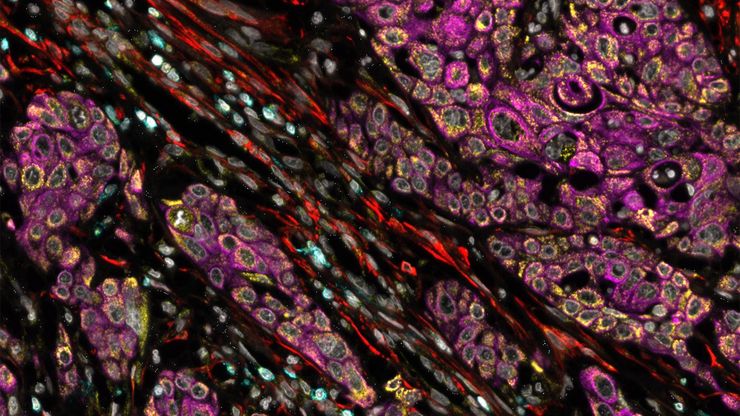
Characterizing tumor environment to reveal insights and spatial resolution
Antibodies from Cell Signaling Technology are validated for use with the Cell DIVE multiplexing workflow and used to probe cell lineages in the tumor microenvironment

Dig Deeper Into the Complexities of Pancreatic Cancer with Multiplex Imaging
Cell DIVE is an iterative staining workflow for multiplexed imaging that unveils biological pathways to dig deeper into the complexities of pancreatic cancer.

Complex Made Simple: Antibodies in Multiplexed Imaging
Build panels, plan studies, and get the most from precious reagents using this antibody multiplexing guide from Leica Microsystems
Cell DIVE: Unraveling Pathways in Pancreatic Cancer
Unlock new insights into tumour-immune interactions with spatial biology. Join our webinar to learn how to label >60 biomarkers per tissue section and use advanced clustering and analysis techniques…

Multiplexed Imaging Types, Benefits and Applications
Multiplexed imaging is an emerging and exciting way to extract information from human tissue samples by visualizing many more biomarkers than traditional microscopy. By observing many biomarkers…
