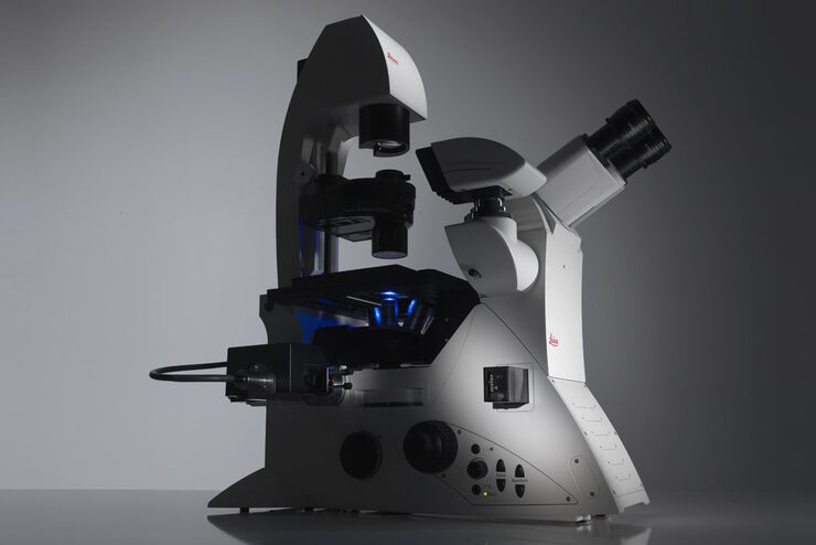
DMi8 S Platform
倒立
光学顕微鏡
製品紹介
Home
Leica Microsystems
DMi8 S Platform 倒立顕微鏡ソリューション
謎を解く鍵をみつけよう
最新の記事を読む
Studying Virus Replication with Fluorescence Microscopy
The results from research on SARS-CoV-2 virus replication kinetics, adaption capabilities, and cytopathology in Vero E6 cells, done with the help of fluorescence microscopy, are described in this…

Epi-Illumination Fluorescence and Reflection-Contrast Microscopy
This article discusses the development of epi-illumination and reflection contrast for fluorescence microscopy concerning life-science applications. Much was done by the Ploem research group…
")
Introduction to Fluorescent Proteins
Overview of fluorescent proteins (FPs) from, red (RFP) to green (GFP) and blue (BFP), with a table showing their relevant spectral characteristics.

Differential Interference Contrast (DIC) Microscopy
This article demonstrates how differential interference contrast (DIC) can be actually better than brightfield illumination when using microscopy to image unstained biological specimens.
 cells taken with phase contrast.")
Phase Contrast and Microscopy
This article explains phase contrast, an optical microscopy technique, which reveals fine details of unstained, transparent specimens that are difficult to see with common brightfield illumination.
Immersion Objectives
How an immersion objective, which has a liquid medium between it and the specimen being observed, helps increase the numerical aperture and microscope resolution is explained in this article.

Precise Spatial Proteomic Information in Tissues
Despite the availability of imaging-based and mass-spectrometry-based methods for spatial proteomics, a key challenge remains connecting images with single-cell-resolution protein abundance…

Studying Wound Healing of Smooth Muscle Cells
This article discusses how wound healing of cultured smooth muscle cells (SMCs) in multiwell plates can be reliably studied over time with less effort using a specially configured Leica inverted…
How to Prepare your Specimen for Immunofluorescence Microscopy
Immunofluorescence (IF) is a powerful method for visualizing intracellular processes, conditions and structures. IF preparations can be analyzed by various microscopy techniques (e.g. CLSM,…
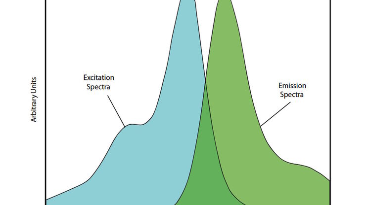
Fluorescent Dyes
A basic principle in fluorescence microscopy is the highly specific visualization of cellular components with the help of a fluorescent agent. This can be a fluorescent protein – for example GFP –…
 and astrocytes (green) in a cortical spheroid derived from human induced pluripotent stem cells.")
Download The Guide to Live Cell Imaging
In life science research, live cell imaging is an indispensable tool to visualize cells in a state as in vivo as possible. This E-book reviews a wide range of important considerations to take to…
Factors to Consider When Selecting a Research Microscope
An optical microscope is often one of the central devices in a life-science research lab. It can be used for various applications which shed light on many scientific questions. Thereby the…
Microscopy in Virology
The coronavirus SARS-CoV-2, causing the Covid-19 disease effects our world in all aspects. Research to find immunization and treatment methods, in other words to fight this virus, gained highest…

Koehler Illumination: A Brief History and a Practical Set Up in Five Easy Steps
In this article, we will look at the history of the technique of Koehler Illumination in addition to how to adjust the components in five easy steps.

Introduction to Mammalian Cell Culture
Mammalian cell culture is one of the basic pillars of life sciences. Without the ability to grow cells in the lab, the fast progress in disciplines like cell biology, immunology, or cancer research…

Introduction to Widefield Microscopy
This article gives an introduction to widefield microscopy, one of the most basic and commonly used microscopy techniques. It also shows the basic differences between widefield and confocal…

Photoactivatable, Photoconvertible, and Photoswitchable Fluorescent Proteins
Fluorescent proteins (FPs) such as GFP, YFP or DsRed are powerful tools to visualize cellular components in living cells. Nevertheless, there are circumstances when classical FPs reach their limits.…
 illuminated with wide-band UV excitation. Note the tissue structure with blue autofluorescence. Right: Same tissue and same immunostaining with FITC label illuminated with epi-illumination using narrow")
Milestones in Incident Light Fluorescence Microscopy
Since the middle of the last century, fluorescence microscopy developed into a bio scientific tool with one of the biggest impacts on our understanding of life. Watching cells and proteins with the…

Chronic Inflammation Under the Microscope
In the course of chronic inflammation certain body areas are recurrently inflamed. This goes along with many human diseases. With the help of widefield light microscopy, the underlying processes can…

Gene Editing with CRISPR/Cas9 - Breakthrough in Genome Engineering
The CRISPR/Cas9 system is one of several different bacterial systems for defense against viral attacks. It consists of two main components. One is a small piece of RNA which binds to the viral target…

Studying Caenorhabditis elegans (C. elegans)
Find out how you can image and study C. elegans roundworm model organisms efficiently with a microscope for developmental biology applications from this article.

Infinity Optical Systems
“Infinity Optics” refers to the concept of a beam path with parallel rays between the objective and the tube lens of a microscope. Flat optical components can be brought into this “Infinity Space”…
Handbook of Optical Filters for Fluorescence Microscopy
Fluorescence microscopy and other light-based applications require optical filters that have demanding spectral and physical characteristics. Often, these characteristics are application-specific and…

Video Tutorial: How to Align the Bulb of a Fluorescence Lamp Housing
The traditional light source for fluorescence excitation is a fluorescence lamp housing with mercury burner. A prerequisite for achieving bright and homogeneous excitation is the correct centering and…

Video Tutorial: How to Change the Bulb of a Fluorescence Lamp Housing
When applying fluorescence microscopy in biological applications, a lamp housing with mercury burner is the most common light source. This video tutorial shows how to change the bulb of a traditional…
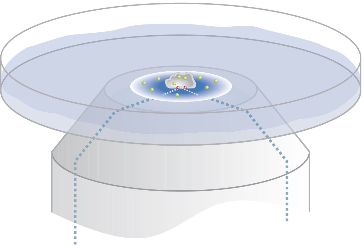
Controlling the TIRF Penetration Depth is Mandatory for Reproducible Results
The main feature of total internal reflection fluorescence (TIRF) microscopy is the employment of an evanescent wave for the excitation of fluorophores instead of using direct light. A property of the…
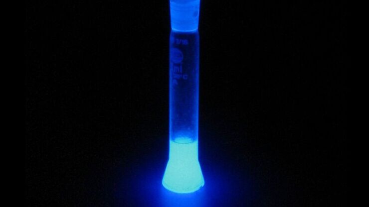
Basic Principles of Luminescence
There are a lot of light-emitting processes occurring in nature. Luminescence is an umbrella term for those kinds of events where light emission is not the result of high temperatures. This article…

Total Internal Reflection Fluorescence (TIRF) Microscopy
Total internal reflection fluorescence (TIRF) is a special technique in fluorescence microscopy developed by Daniel Axelrod at the University of Michigan, Ann Arbor in the early 1980s. TIRF microscopy…

Fluorescent Proteins - From the Beginnings to the Nobel Prize
Fluorescent proteins are the fundament of recent fluorescence microscopy and its modern applications. Their discovery and consequent development was one of the most exciting innovations for life…
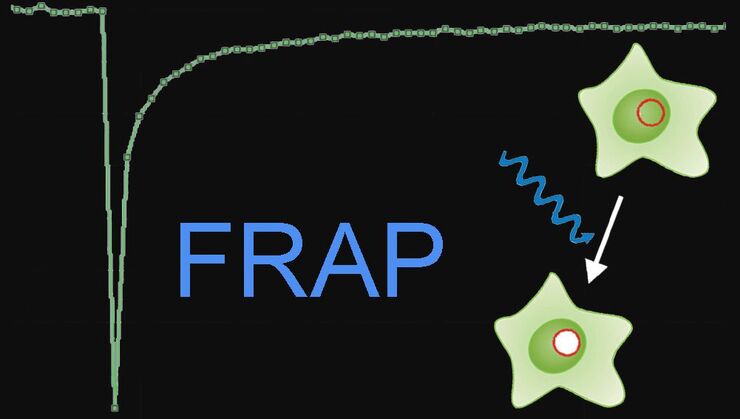
Fluorescence Recovery after Photobleaching (FRAP) and its Offspring
FRAP (Fluorescence recovery after photobleaching) can be used to study cellular protein dynamics: For visualization the protein of interest is fused to a fluorescent protein or a fluorescent dye. A…

An Introduction to Fluorescence
This article gives an introduction to fluorescence and photoluminescence, which includes phosphorescence, explains the basic theory behind them, and how fluorescence is used for microscopy.

Optical Contrast Methods
Optical contrast methods give the potential to easily examine living and colorless specimens. Different microscopic techniques aim to change phase shifts caused by the interaction of light with the…

Integrated Modulation Contrast (IMC)
Hoffman modulation contrast has established itself as a standard for the observation of unstained, low-contrast biological specimens. The integration of the modulator in the beam path of themodern…

Fluorescence in Microscopy
Fluorescence microscopy is a special form of light microscopy. It uses the ability of fluorochromes to emit light after being excited with light of a certain wavelength. Proteins of interest can be…
応用分野
マイクロマニピュレーション
ライカ マイクロシステムズの簡単で便利なシステムが実現する、自動化された高速のマイクロマニピュレーションを実体験してください。リサーチ用倒立顕微鏡と電子マイクロマニピュレーターを完全装備することで、これらの完全ソリューションは確実な操作、システム振動の低減、そしてルーチンワークおよびトレーニングの双方における時間節約を実現します。
光子操作
光子操作という用語は、事象を引き起こし、長時間にわたり生細胞におけるダイナミックで複雑な挙動を観察するため蛍光分子の性質を利用するための広範な技術を指すものです。ブリーチングであろうと活性化であろうと、変換、アブレーションあるいは結合技術であろうと、研究者は高解像度で事象を実現させ観察するに充分なシステムを必要とします。
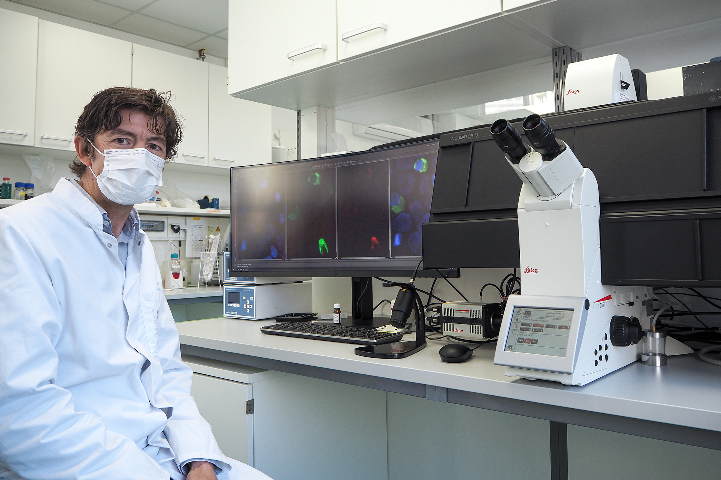
生細胞イメージング
視点を単体の顕微鏡コンポーネントから必要なすべての機能を備えた生細胞イメージングソリューションへと移し、ライカ マイクロシステムズは顕微鏡、LAS X イメージングソフトウェア、カメラおよび専用サードパーティコンポーネントを 1 つの完全な生細胞イメージングシステムに統合します。
ウイルス学
ウイルス研究のためのイメージングと試料作製ソリューション
顕微鏡の高度な技術
高度な顕微鏡技術には、高解像度および超解像のイメージング技術が含まれます。これらの技術は主に、細胞や組織などの試料にできるだけ優しく、極めて高い解像度で生物学的事象を可視化するために使用されます。 研究者は、高度な顕微鏡技術によって、生物学的経路、遺伝子やタンパク質の発現、病気のメカニズムなどに大きな影響を与える生体分子を調べ、理解することができます。
顕微鏡の基本的な技術
顕微鏡の基本的な技術は、顕微鏡ステージ上の試料全体が光源にさらされる形で使用されます。試料全体は、上方(倒立型)または下方(標準的な正立顕微鏡の場合)から白色光で照らされます。試料の透明度や観察対象・観察部位に応じて、複雑な細胞構造を識別するために異なる蛍光粒子を使用します。
暗視野顕微鏡
暗視野コントラスト法は、生体試料の構造や物質試料の不均一な特徴から生じる光の回折や散乱を利用する方法です。
位相差顕微鏡
位相差顕微鏡では、染色することなく、多くの種類の生物試料の構造をより優れたコントラストで観察できます。
微分干渉(DIC)顕微鏡
微分干渉(DIC)顕微鏡は、光源と集光レンズの間に偏光フィルターとウォラストンプリズムを配置した広視野顕微鏡で、対物レンズとカメラセンサーや接眼レンズの間にも偏光フィルターとウォラストンプリズムを配置しています。
もっと知りたいですか?
お気軽にお問合せください
ライカまでお気軽にご相談ください Show local contacts