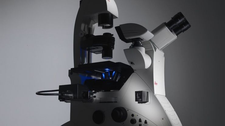
Factors to Consider When Selecting a Research Microscope
An optical microscope is often one of the central devices in a life-science research lab. It can be used for various applications which shed light on many scientific questions. Thereby the…
Faster & Deeper Insights into Organoid and Spheroid Models
Gain deeper, more translatable, insights into organoid and spheroid models for drug discovery and disease research by overcoming key imaging challenges. In this eBook, explore advanced microscopy…
A Guide to C. elegans Research – Working with Nematodes
Efficient microscopy techniques for C. elegans research are outlined in this guide. As a widely used model organism with about 70% gene homology to humans, the nematode Caenorhabditis elegans (also…
Improving Zebrafish-Embryo Screening with Fast, High-Contrast Imaging
Discover from this article how screening of transgenic zebrafish embryos is boosted with high-speed, high-contrast imaging using the DM6 B microscope, ensuring accurate targeting for developmental…
Unlocking the Secrets of Organoid Models in Biomedical Research
Get ready to delve deeper into the world of organoids and 3D models, which are essential tools for advancing our understanding of human health. Navigating these complex structures and obtaining clear…
Designing the Future with Novel and Scalable Stem Cell Culture
Visionary biotech start-up Uncommon Bio is tackling one of the world’s biggest health challenges: food sustainability. In this webinar, Stem Cell Scientist Samuel East shows how they make stem cell…
神経細胞移動の分子的秘密を解き明かす
発達中の脳における特定部位への神経細胞の移動を調べるには、さまざまなアプローチを用いることができます。このウェビナーでは、オックスフォード大学の専門家が、神経発達過程における大脳皮質の機能層への神経細胞移動の分子メカニズムを解明するために使用している顕微鏡ツールとアッセイについて紹介します。これらのプロセスを理解することは、健全な脳の発達の理解を深め、神経発達障害の治療法を改善する可能性につながり…
How Efficient is your 3D Organoid Imaging and Analysis Workflow?
Organoid models have transformed life science research but optimizing image analysis protocols remains a key challenge. This webinar explores a streamlined workflow for organoid research, starting…
Imaging Organoid Models to Investigate Brain Health
Imaging human brain organoid models to study the phenotypes of specialized brain cells called microglia, and the potential applications of these organoid models in health and disease.


