Inspire your research
Super-resolution live cell imaging in multicolor SP8 LIGHTNING
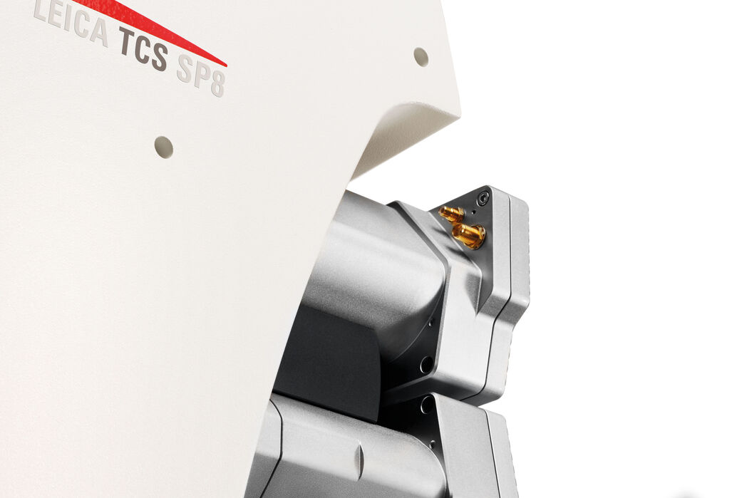
The SP8 LIGHTNING confocal microscope allows you to make proper and detailed observations of fast biological processes. Your experimental work will have the benefit of super-resolution, high-speed imaging, and the capability to image multiple fluorescent markers simultaneously.
Thanks to the exclusive detection concept of LIGHTNING, you can trace the dynamics of multiple molecules, even those expressed at low levels, simultaneously over long recording times in living specimens.
The SP8 LIGHTNING confocal microscope offers you the following performance advantages
- truly simultaneous multicolor imaging in super-resolution down to 120 nm
- live specimen imaging thanks to fast acquisition rates
- sample protection thanks to low phototoxicity
The SP8 LIGHTNING confocal microscope opens up unique experimental options to inspire your research!
Cellular dynamics and details revealed
Extracting the dynamics of cellular and molecular interactions requires the acquisition of multiple fluorescence signals.
The SP8 LIGHTNING confocal system offers you a superior solution to capture up to five true color channels simultaneously at high speed without need for trade-offs in multicolor experiments.
Mouse Colon. Role study of chemokine receptor CX3CR1 in intestinal inflammatory diseases. Green: GFP-CX3CR1, macrophages. B&W: AF 647-CD31, vasculature. Red: PE-CD49B, blood platelets. 15 fps, 512x512 , xyzt, 375 x 375 µm, z: 1601.6 µm. Duration 3 min 197s. Objective 25x0.95 W Fluotar VISIR. Video: Courtesy of W.Y. Lee, Calvin, Phoebe & Joan Snyder Institute for Chronic Diseases, Cumming School of Medicine, University of Calgary, Canada.
Host pathogen interaction in plant cells. Prestudy: Simultaneous monitoring of multiple organelle markers in leaf of transgenic Arabidopsis thaliana plants, 8 fps, 1024x1024, PL APO 40x 1.1 W. Cyan: mTurquoise2, nuclear localization site (N7). Green: Venus, microtubules (MAP4). B&W: tagRFP-T, peroxisomes (SKL). Red: mKate2, plasma membrane (LTI6b). Courtesy of Dr. Hasan Ghareeb, Prof. Dr. Volker Lipka, Department of Plant Cell Biology, University of Göttingen, Germany.
Super-resolution in real time
Frame rates for recording processes in living samples can be adjusted by FOV scanning systems
Benefit from recording various physiological processes at very high rates up to 40 fps at 512x512 offered by resonant scanning.
Process your data with LIGHTNING in real time to obtain images with super-resolution, down to 120 nm, enabling you to see the finest details in your living specimens.
Read more in Science Lab
Obtain more information from your experiment
Get images with a large field of view showing super-resolved structures and allowing the observation of molecular interactions and dynamics.
The SP8 LIGHTNING allows you to perform proper sampling over a specimen area at least six times larger than with any other confocal system. It achieves this level of performance using high-speed resonant scanning with imaging formats of up to 2496 x 2496 pixels.
HeLa Kyoto cells. P24-EYFP, tubulin-SiR dye. 3D time lapse series. Combination of highest speed and resolution, using a PL APO 63x/1.2 W objective, pixel size of 53nm, format 1000 x 400 pixels at 32 fps. Sample courtesy of Dr. Juan Jung, EMBL, Heidelberg
Upper movie: Confocal raw data, no averaging, simultaneous acquistion. Lower movie: Super-resolved 3D time-lapse imaging powered by LIGHTNING detection.
Enhanced signal-to-noise ratio
The SP8 LIGHTNING confocal system takes the image acquisition speed and signal-to-noise ratio to a new level, both for standard and resonant scanning.
Focus on the field of interest: acquisition speed can be greatly improved by using freely tunable scan formats and rotation.
Benefit from the full recording speed as LIGHTNING enables acquisition with a minimum of averaging.
Specimens protected for long imaging times
Designed for sensitivity, the SP8 LIGHTNING confocal system allows you to follow dynamic processes in organisms over long periods of time without disturbing their development. Lowest illumination intensity greatly reduces phototoxicity.
The SP8 LIGHTNING makes sure every photon counts. Its detection concept provides imaging with enhanced sensitivity and the highest efficiency. This optimization is possible due to the synergy of super-sensitive hybrid detection integrated into a filter-free prism-based spectral detection system.
Zebrafish embryo, development of the lateral line organ. 3D time-lapse using the 8 kHz resonant scanner and a PL APO 40x 1.1 W objective. Image format 1024x512, duration 8 h 28 min. Green: Membranes, GFP, red: Nuclei, tdTomato. Courtesy of Jonas Hartmann, Gilmour Group, EMBL Heidelberg, Germany.
NAVIGATOR tile scan (3x2) of a whole mount of mouse diaphragm muscle. Blue: Nuclei, B&W: F-actin, Red: Nicotinic acetylcholine receptors, a principal postsynaptic component of neuromuscular junctions, Green: Tyrosine hydroxylase, a marker for sympathetic neurons. LIGHTNING automatically reveals the finest details applying an adaptive process for extraction of hidden information in the image. Sample courtesy of Tatjana Straka and Rüdiger Rudolf, University of Applied Science, Mannheim, Germany.
Navigate your specimen with ease
The LAS X Navigator software allows for easy correlation of high resolution images with slide overviews, even created with a mobile phone.
Define with a single mouse click the slide areas to be scanned. Use a fast mosaic scan to get a detailed view of the defined area where you can zoom into your region of interest.
The correlation includes not only intensity information, but also quantitative data like excitation spectra and fluorescence lifetime imaging.
With the LAS X Navigator, you can use a large selection of sample carrier templates to do easy multiwell plate screening. Create a quick overview using the spiral scan in each well and use the auto-focus map to keep your focus in positon. Quantify image data generated by random position or selected well imaging.
Fast track to results
The workflow-oriented design of the LAS X and intuitive software wizards guide you step-by-step through image acquisition, processing, and analysis. Tailor LAS X to your needs with additional software packages, like 3D Visualization and 3D Analysis, which allow you to understand the topology of your 3D image and quantify various aspects of intracellular structures. The examination and analysis of your experiments become straightforward with the SP8 LIGHTNING system and LAS X software.
LAS X Live Data Mode
- Easy setup of complex, interactive time-lapse experiments
- Change hardware parameters during a running experiment for maximum control
- Trigger functions to synchronize image acquisition with external devices
LAS X MicroLab
- Wizards for user friendly setup and analysis of FRAP, FLIP and FRET
- Optimized for fast recording speeds
- User guidance for minimal learning effort
LAS X 3D Visualization
- Fast reconstruction and processing of 3D data by GPU-based algorithms
- Interactive shadow projection to emphasize three dimensional structures
- Powerful clipping tool for channel specific user defined segmentations
- Sophisticated Movie Editor for the generation of complex 3D animationsU: LAS X microscope software with additional software packages
Be inspired with multiple imaging modalities
In whichever direction your research will evolve, the SP8 LIGHTNING confocal microscope is open to adapt to your scientific requirements.
You can combine and accommodate different research methods in the SP8 LIGHTNING ranging from confocal microscopy with super-resolution to STED nanoscopy.
Be free to add methods such as multiphoton microscopy, lifetime imaging, light sheet imaging or CARS depending on your research question.
Let the SP8 LIGHTNING inspire your research.
Interested to know more?
Talk to our experts. We are happy to answer all your questions and concerns.
Contact UsDo you prefer personal consulting?
-
Leica Microsystems Inc.1700 Leider LaneBuffalo Grove, IL 60089 United StatesOffice Phone : +1 800 248 0123Service Phone : 1 800 248 0223Fax : +1 847-236-3009
-
JH Technologies, Inc.213 Hammond AvenueFremont, 94539 United StatesOffice Phone : 408-436-6336Fax : 408-436-6343
-
Precision Lens5715 West Old Shakopee Road Unit 150Bloomington, 55437 United StatesOffice Phone : 952-881-0414
-
D.M.I. Medical Inc.Life Science Research: Confocal Microscopy, Widefield, Nanotechnology, Industry Microscopy, Clinical Microscopy, Surgical Microscopy, Digital Pathology, Cytovision, Aperio, Ariol, Histology Instruments, Histology Consumables, Advanced Staining Instruments, Advanced Staining Consumables, Kreatech Reagents, Thermobrite / Thermobrite Elite4611 S. University Dr. Suite 435Davie, 33328 United StatesOffice Phone : +1-954-538-3808Service Phone : +1-954-328-0693Fax : +1-866-492-6373
-
D.M.I. Medical IncHistology Systems, Novocastra Reagents and associated instruments, EM Sample Preparation4611 S. University Dr. Suite 435Davie, 33328 United StatesOffice Phone : +1-954-538-3808Service Phone : +1-954-328-0693Fax : +1-866-492-6373
-
ECLIPSE SURGICAL SUPPLY LLC616 FAIRWAY MIST CTMINT HILL, 28227 United StatesOffice Phone : 410-299-2711
-
Great Plains Surgical, Inc.3605 Hidden CircleWest Fargo, 58078 United StatesOffice Phone : 701-391-3333Service Phone : 701-391-3333Fax : 701-364-0307
-
ICMAS Inc.120 McCammon AveMaryville, 37804 United StatesOffice Phone : 865-984-8058
-
Integrity Surgical2303 Ranch Road 620 S Suite 160-310Lakeway, 78734 United StatesOffice Phone : 713-201-8787
-
Next Case Surgical943 Maple Avenue - Upper LevelDowners Grove, 60515 United StatesOffice Phone : 312-520-7600Fax : 219-662-7353
-
Proficient Surgical Equipment, Inc99 Seaview Blvd. Suite CPort Washington, 11050 United StatesOffice Phone : 516-487-1175Fax : 516-487-1191
-
JH Technologies, Inc. - Neast213 Hammond AveFremont, 94539 United StatesOffice Phone : 408-436-6336Fax : 408-436-6343
-
JH Technologies So. Cal213 Hammond AveFremont, 94539 United StatesOffice Phone : 408-436-6336Fax : 408-439-6343
-
Q Surgical28 Liberty Ship Way Suite 2830Sausalito, 94965 United StatesOffice Phone : 415-331-1020Fax : 415-462-5186
-
Rohlf Medical, Inc.2600 S. Minnesota Ave Suite 101Sioux Falls, 57105 United StatesOffice Phone : 605-334-8655Fax : 605-334-9102
-
Shawnee Medical Products, Inc.8401 Juniper LanePrairie Village, 66207 United StatesOffice Phone : 913-341-7520Fax : 913-341-2318
-
Spective, LLC3103 Glenwood DriveDurham, 27705 United StatesOffice Phone : 9194526288
-
Spective LLC - Canada3103 Glenwood DriveDurham, 27705 United StatesOffice Phone : 9194526288
-
SurgicalOne, Inc.23030 E Industrial DriveSAINT CLAIR SHORES, 48080 United StatesOffice Phone : 586-775-3300Fax : 586-775-3301
-
TAP Medical Products, Inc.4580 Klahanie Drive S.E. #452Issaquah, 98029 United StatesOffice Phone : 425-785-9993Fax : 800-832-1859
-
Western Microsurgical6721 Esperanza DriveCastle Rock, 80108 United StatesOffice Phone : 720-244-4679Fax : 303-331-8128
-
NCI, Inc.7125 Northland Terrace N. Suite 100Brooklyn Park, 55428 United StatesOffice Phone : 763-559-3008Fax : 763-559-7372
-
NCI, Inc.7125 Northland Terrace N. Suite 100Brooklyn Park, 55428 United StatesOffice Phone : 763-559-3008Fax : 763-559-7372
-
W. Nuhsbaum Inc.760 Ridgeview DriveMcHenry, 60050 United StatesOffice Phone : 815-385-5200Fax : 815-385-5236
-
Angstrom Scientific Inc.120 N CENTRAL AVE STE 3Ramsey, 07446 United StatesOffice Phone : 2019627222Fax : 2019628290
-
Associated Microscope Inc.412 Holly Hill LnBurlington, 27215 United StatesOffice Phone : 336-538-1842Fax : 336-538-1847
-
I. Miller Precision Optical Instruments Inc.325 Bustleton PikeFeasterville Trevose, 19053 United StatesOffice Phone : 215-925-2285Fax : 215-925-8374
-
Marathon Medical, Inc.1408 Christian Avenue Suite 35Durham, 27705 United StatesOffice Phone : 919-309-4463Service Phone : 919-812-9500Fax : 919-309-4742
-
DB Surgical12480 West Atlantic Blvd Suite 1Coral Springs, 33071 United StatesOffice Phone : 954-340-2727Fax : 954-340-1221