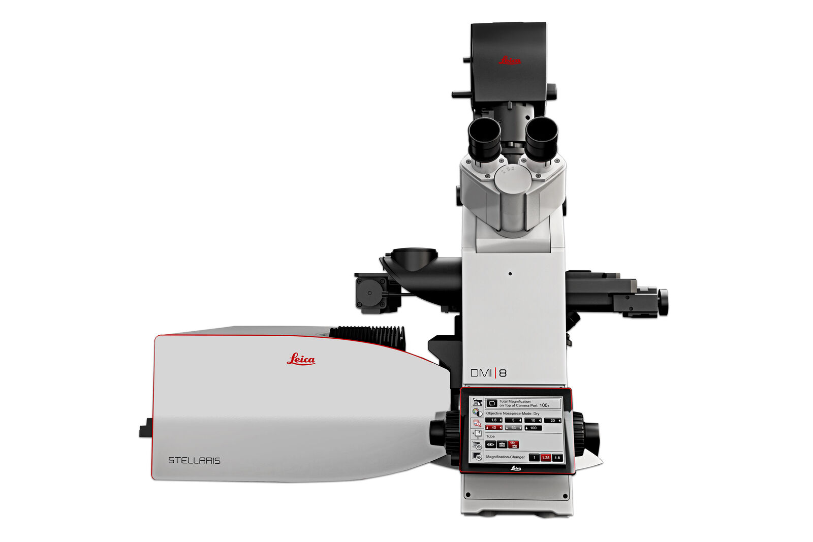
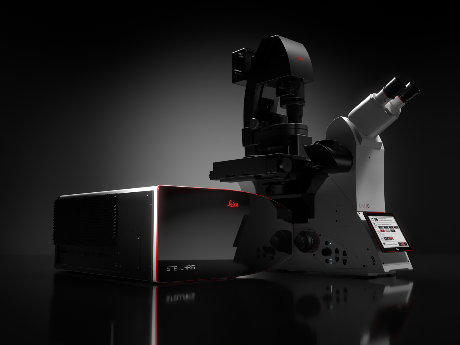
STELLARIS Confocal Microscope Platform
Next generation STELLARIS is a confocal microscope platform designed around your research needs.
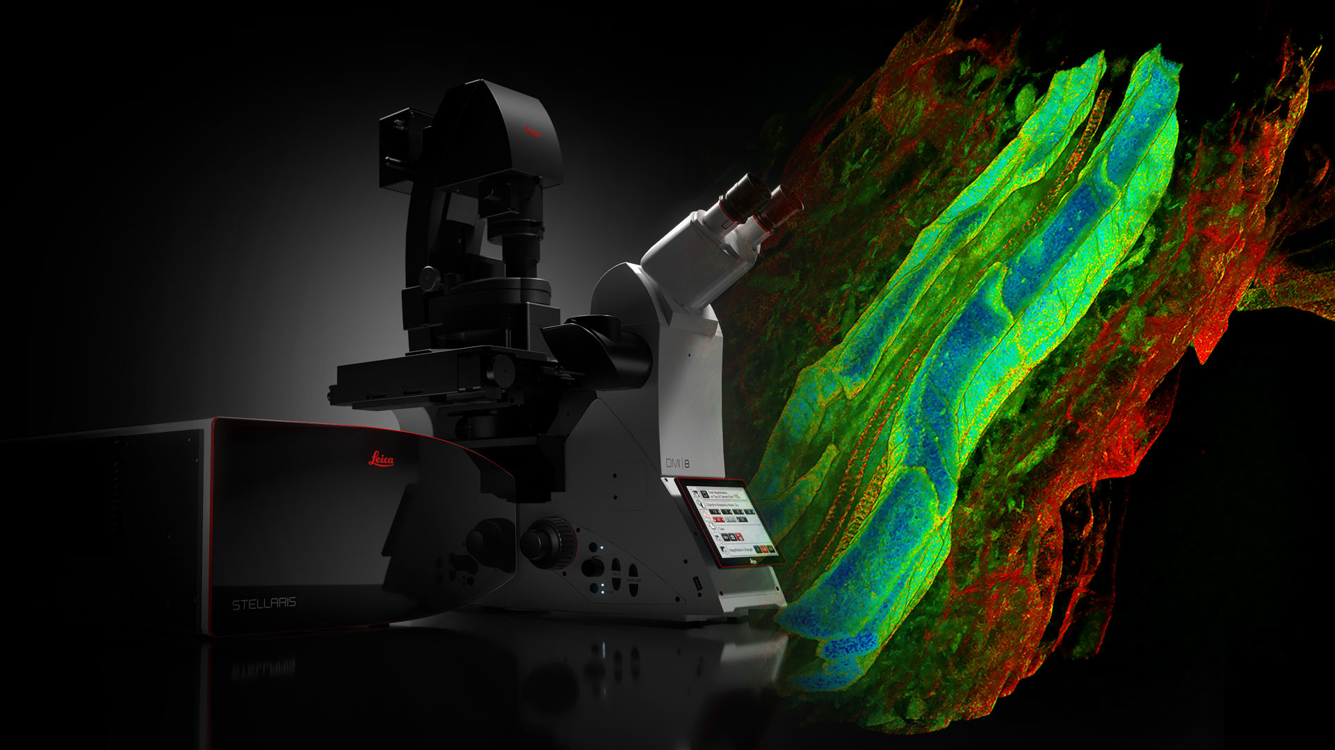
STELLARIS confocal microscopes can be combined with all Leica modalities, including FLIM, STED, MP, DLS, and CRS. The next generation STELLARIS confocal platform is your fast track to rewarding research. Power. Potential. Productivity.
Popular Configurations

STELLARIS Personal Confocal MicroscopePersonal Confocal Microscope STELLARIS is a true confocal point scanning system with beam splitters and has Spectral Detection, Photon Counting Detectors, and Confocal Super Resolution! STELLARIS confocal microscope also offers the following modalities, including Autonomous Microscopy, Digital Light Sheet, Dynamic Signal Enhancement, and more. Include: A fully equipped confocal microscope with 3 Power HyD S detectors, 3 VIS Laser Lines, 405 nm Laser, Motorized X-Y Stage, and Confocal LIGHTNING Super Resolution package - Request a Quote Today!* An array of key features that facilitates imaging: - 3 VIS Laser Lines and a 405 nm Laser - 3 Photon Counting Detectors - Motorized X-Y Stage - Confocal LIGHTNING Super Resolution package - Upgradable to additional detectors (up to a total of 5) or additional lasers etc. in the future
Loading...
View product details+
Product includes
Loading...
|
||||||||||||||||||||||||||||||||||||||||||||||||||||||||||||||||||||||||||||||||||||||||||||||||||||||||||||||||||||||||||||||||||||

Don’t see your option? Request for individual Quote
Show popular configurations
Explore and buy pre-configured microscopy solutions in our online shop. Enjoy a seamless online shopping experience.
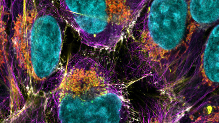
The Power to see more
The Power HyD detector family provides higher photon detection efficiency (PDE)*, extremely low dark noise, and sensitive spectral detection from 410 to 850 nm.
- Enhanced image quality: Ideal combination of brightness, resolution and contrast.
- Complete spectral freedom: Our next generation White Light Lasers allow you to simultaneously use up to 8 single excitation lines from across the spectrum. Image more fluorophore combinations than with any other confocal platform and use more labels in parallel, expanding your options into the NIR range.
- Gentle live cell imaging: Preserve your sample’s integrity and image for longer periods of time through efficient signal acquisition at the lowest required levels of illumination
*2x higher Photon Detection Efficiency (PDE) compared to conventional multi-alkali Photomultiplier Tubes (PMT) and 3x higher in the extended red range.
The Potential to discover more
STELLARIS unique TauSense technology allows you to extract an extra layer of information from every sample and increase the scientific impact of your research. TauSense is comprised of application-oriented imaging tools based on fluorescence lifetime that you can use to explore the function of molecules within the cellular context.
- TauContrast provides immediate access to functional information, such as metabolic status, pH, and ion concentration.
- TauGating improves the quality of your images by removing unwanted fluorescence contributions.
- TauSeparation helps you expand the combination of fluorescent signals in your experiment beyond the spectral options.
- TauInteraction provides straightforward detection and quantification of molecular interactions (e.g. protein-protein interaction).
The Productivity to do more
Streamlined setup and acquisition: ImageCompass, the STELLARIS smart user interface, provides users with an easy and intuitive way to set up even the most complex experiments with just a few clicks.
- Simply fast: Superb image quality in real time and at full speed with the LIGHTNING super-resolution, Dynamic Signal Enhancement (DSE) powered by Aivia.
- Intuitive User Interface: ImageCompass guides you from the experiment setup through acquisition.
- Optimize your experiment: Tools such as LAS X Navigator are seamlessly integrated for straightforward imaging.
Access data that matter
Get high-quality results faster with Autonomous Microscopy powered by Aivia.
- The AI-based rare event detection workflow for life science on STELLARIS autonomously detects up to 90 % of rare events in biological samples.
- Radically shorten the data acquisition time by up to 70 %, as objects of interest are exclusively identified and recorded. Record only what you need to, saving valuable space on your hard drive.
- Spend less time at the microscope: The autonomous rare event detection workflow requires only a fraction of the time you would usually need.
- Conduct experiments that were previously not possible due to time constraints and complexity with Autonomous Microscopy powered by Aivia.
STELLARIS with WLL
STELLARIS the only confocal system with an integrated WLL, combined with our proprietary Acousto-Optical Beam Splitter (AOBS) and the Power HyD detector family. Together with the unique TauSense technology, STELLARIS sets a new standard for the quality of images and quantity of information generated.
STELLARIS with White Light Laser can be combined with FAst Lifetime CONtrast (FALCON), STED, Deep In Vivo Explorer (DIVE), Digital Light Sheet (DLS), Cryo and CARS. STELLARIS maximizes the potential of these modalities and give you the power and potential to set new standards for research.
STELLARIS
STELLARIS offers Spectral Detection, Photon Counting Detectors, and Confocal Super Resolution.
Multi-channel confocal imaging is easily attainable thanks to the smart user interface, ImageCompass, which guides you intuitively through your experiment set up and acquisition.