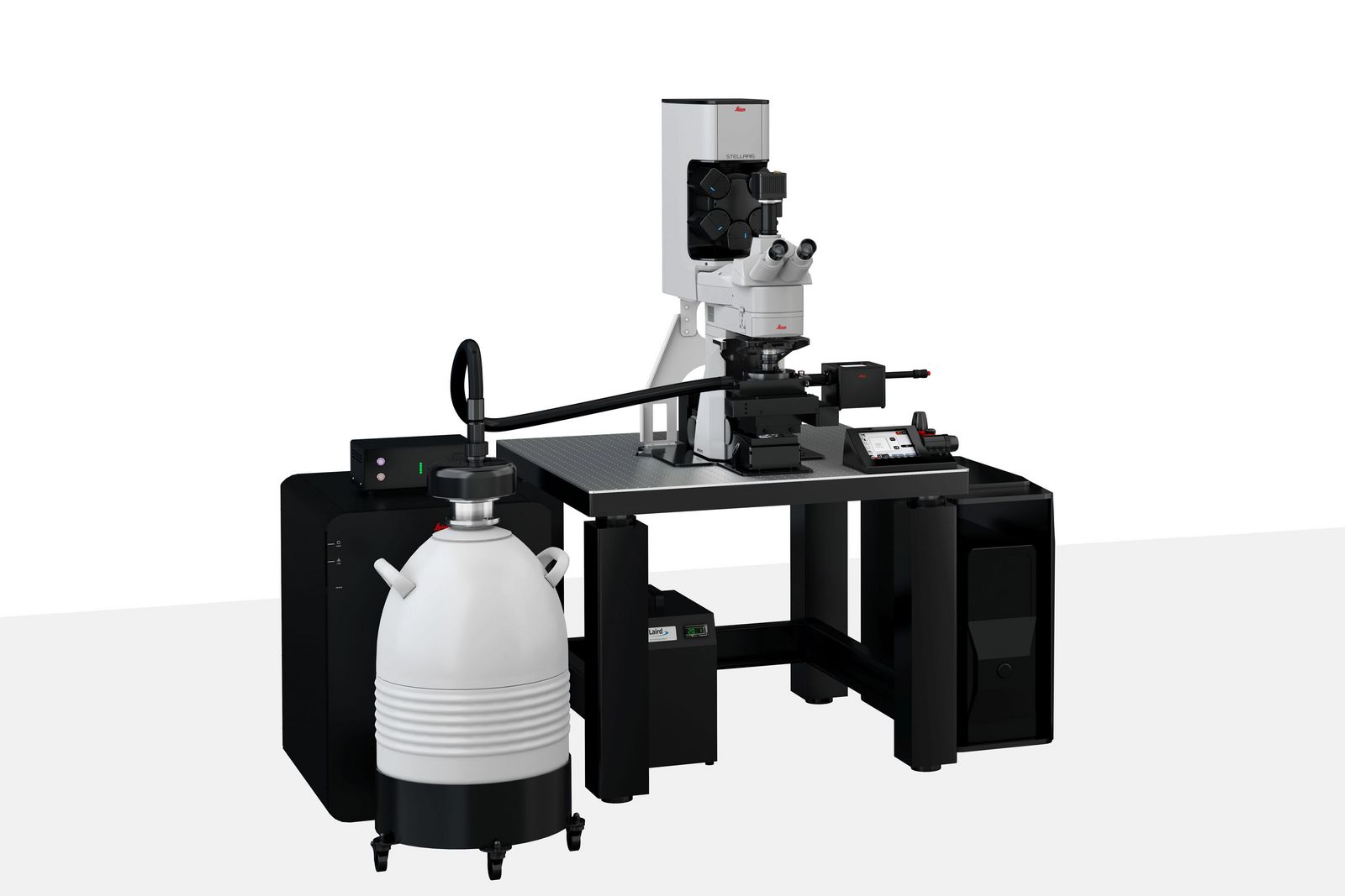
STELLARIS Cryo Confocal Microscope
STELLARIS Cryo is a confocal microscope system that helps you to target your area of interest for cryo-electron tomography (CryoET). The STELLARIS Cryo gives you the precision to target reliably, while offering superior performance you can count on and sample safety for your experiments.
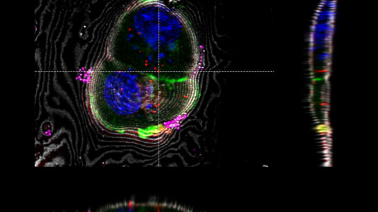
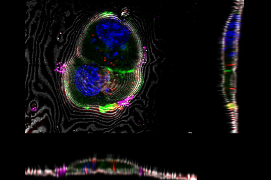
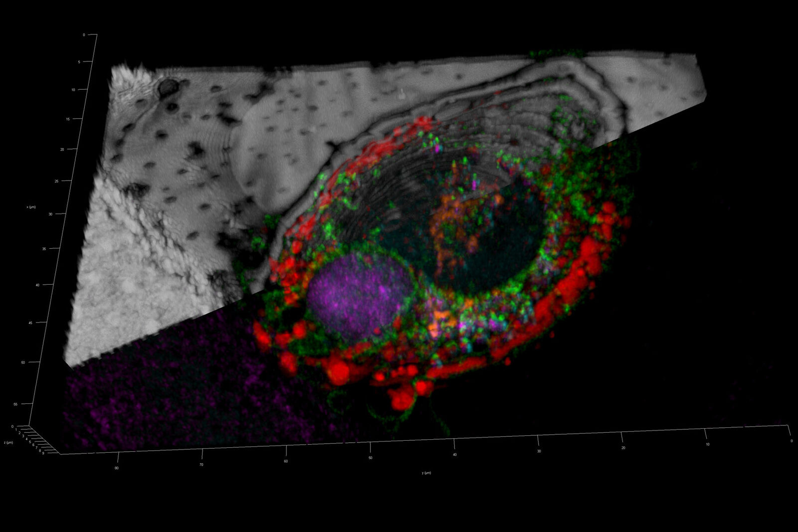
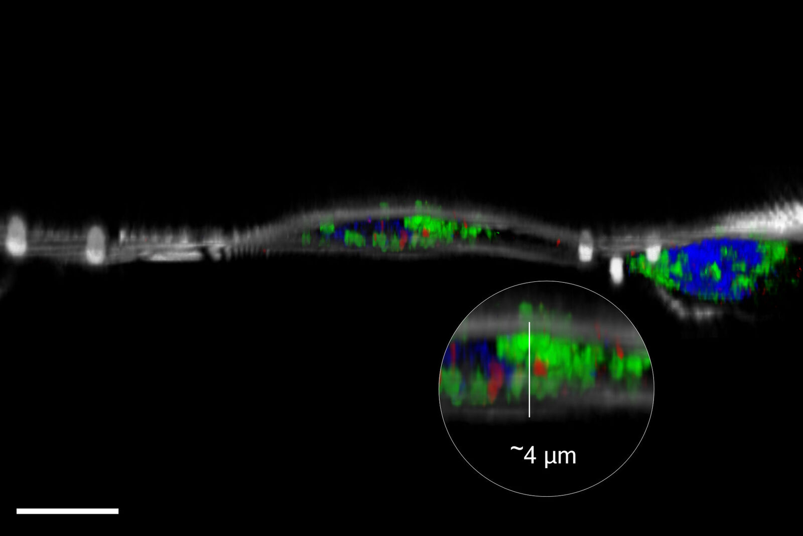
Resolve and identify intracellular targets in 3D
For correlative workflows, it is crucial to retrieve the exact target coordinates identified in the cryo microscope because fluorescence signals are not visible in subsequent electron microscope steps.
Our LAS X Coral Cryo software allows for precise positioning of open format coordinate markers in a nanometer scale. Different marker types serving different functions are also available for landmark, lamellae and bead marking. STELLARIS Cryo with LAS X Coral Cryo software provides 3D confocal data in open file formats that can be imported to any EM software: TIFF, LIFE, ome.tiff.
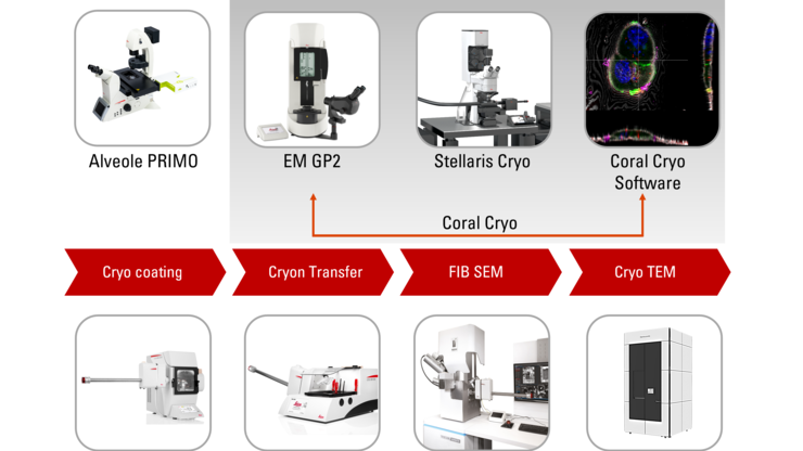
Reduce handling steps between STELLARIS cryo confocal and cryo-FIB.
The dedicated Coral Cryo cryo-electron tomography workflow ensures sample viability, quality checks, and most of all, a precise and reliable 3D imaging.
STELLARIS Cryo, including cryo stage and shuttle, is designed to be used in conjunction with LAS X Coral Cryo software, as well as a variety of seamless integration and transfer options to cryo FIB or VCT stages. New cryo stage increases working range in non-cryo applications, expanding STELLARIS usability for confocal imaging.
You can keep the sample safe during the transfer as all components are compatible.

New generation STELLARIS
Observe finer details in every sample, capture faintest signals, and gather more precise and reliable data across the spectrum.
Groundbreaking discoveries become possible by incorporating an extra dimension into your experiments with fluorescence lifetime-based information.
Enhanced productivity comes with simplified setup and navigation.
Learn more about the STELLARIS Confocal Platform.
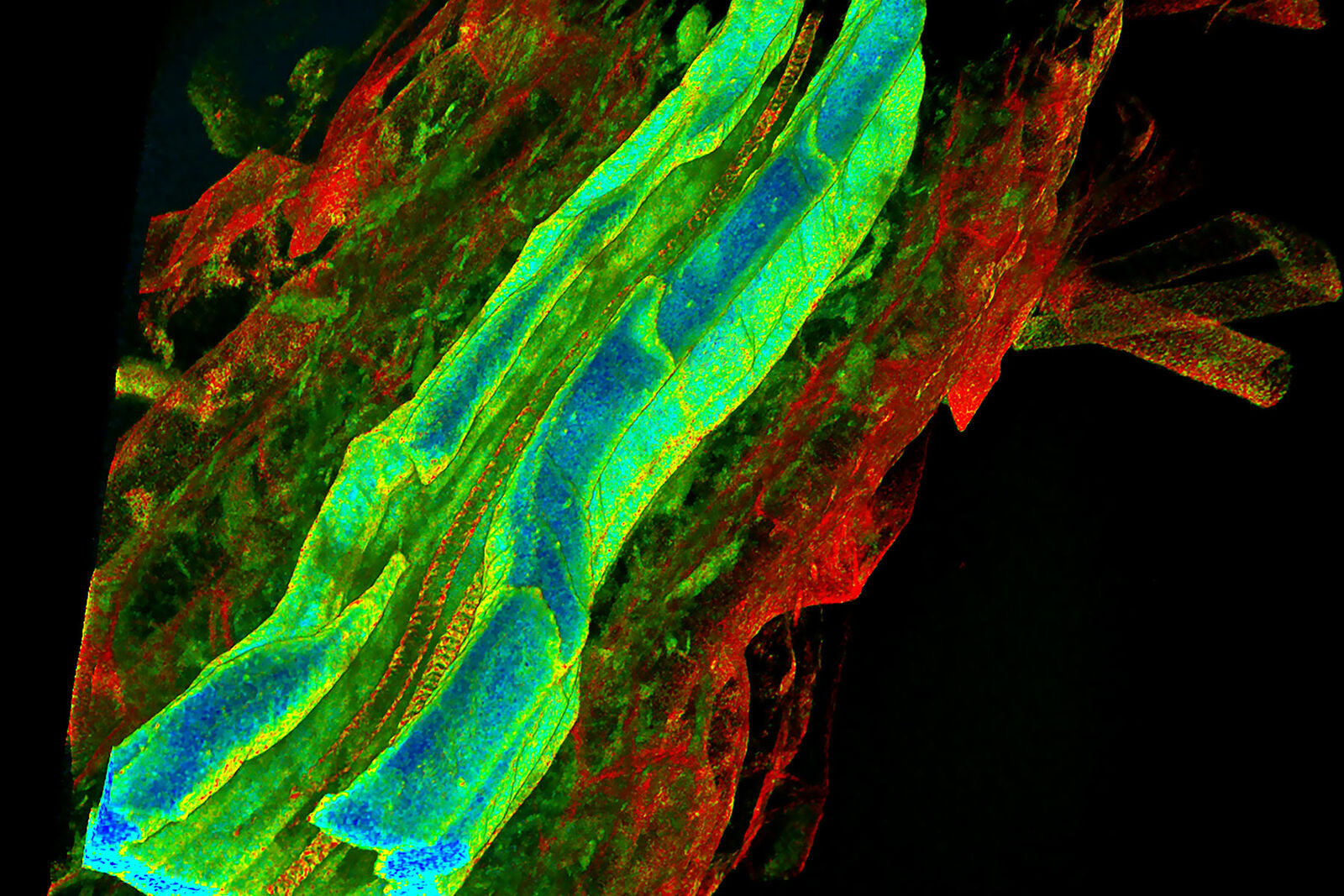
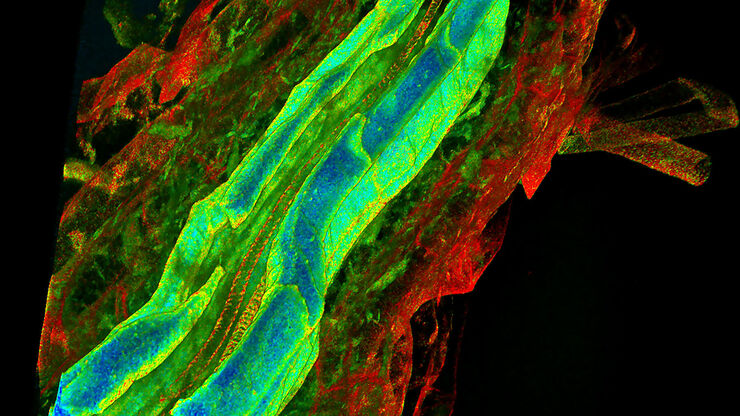
Differentiate targets from background with advanced imaging
The perfect synergy between our Power HyD detectors family, fully optimized beam path, and next-generation White Light Lasers delivers exceptional imaging performance. Your results are clearer, with greater detail derived from brighter signals, more contrast, and increased sensitivity – even when imaging low-abundance labels and events.
Maximize the information you extract from your vitrified specimen and get in-depth answers to scientific questions with the unique LIGHTNING detection concept, an adaptive extraction process that reveals fine structures and details in the image information.
This allows you to target 3D structures of interest more precisely: in room temperature and under cryogenic conditions, preparing for the next steps in your EM microscopy workflow.
Performance you can count on
In cryo-electron microscopy, monitoring ice thickness throughout the workflow is crucial, and the earlier it is checked and assessed, the better. If the ice is too thick, it can cause issues during the milling stage, while if it is too thin, it may affect the stability of the sample.
STELLARIS Cryo offers two modes for checking ice thickness: a fast camera overview and a confocal reflection mode, which determines the ice thickness in 3D.
The Cryo CLEM objective HC PL APO 50x/0.90 has been specifically developed to meet the needs of cryo microscopy, providing the advantages of super-resolution without the need for liquid immersion.
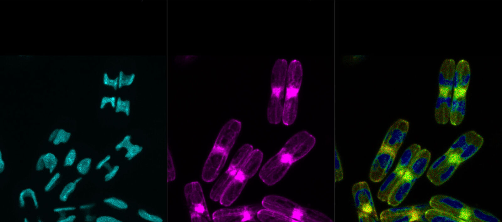
Remove unwanted signal using lifetime-based information
STELLARIS has re-imagined confocal microscopy by providing you an extra dimension of information in every experiment. With TauSense, we put revolutionary new lifetime-based tools at your fingertips. With just a few clicks, you can remove unwanted signal to reveal more detail in your images, multiplex more labels in a single experiment, separate fluorophores that have overlapping spectra, and apply the power of lifetime-based information to explore localized functional and micro-environmental changes. With TauSense, STELLARIS opens a world of new possibilities for your research.
TauSense in STELLARIS Cryo allows you to exploit possible difference in fluorescence lifetime to separate overlapping fluorophores. TauSense can also be used to identify unwanted signal contributions such as grid reflection.
Minimize contamination during imaging
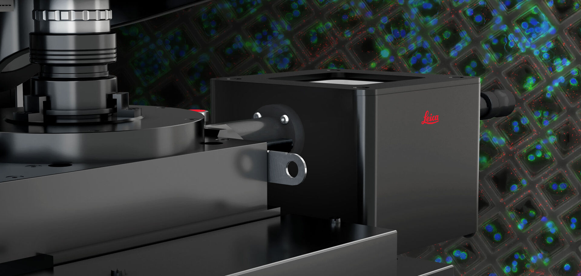
Throughout cryo workflows, samples must be maintained under dehumidified and cryogenic conditions immediately after vitrification. The Cryo Microscopy Kit which includes a cryo stage and shuttle, ensures safe and intuitive sample loading as well as straightforward transfer, all while maintaining the sample under optimal cryo conditions.
The cryo stage allows you to perform light microscopy under stable cryogenic conditions by maintaining a constant overpressure of gaseous nitrogen against the surrounding atmosphere. The stage supports all necessary movements for high-resolution 3D imaging.
To enhance the safety of the loading process, an optional cover box that houses the transfer shuttle is available. This cover box ensures safe loading in all conditions, provides additional process control, and extends the safe loading time window.