STELLARIS Cryo
Confocal Microscopes
Products
Home
Leica Microsystems
STELLARIS Cryo Confocal Microscope
Target what matters for your cryo-tomography workflow
Read our latest articles
 labeled with membrane-permeable calcein, high-pressure frozen in salt water using EM ICE.")
High-Pressure Freezing for Organoids: Cryo CLEM & FIB Lift Out
Master cryo EM workflow steps for challenging 3D samples: when to choose HPF vs. plunge freezing, reproducible blotting/ice control, contamination aware transfers, Cryo CLEM 3D targeting in organoids,…

From Bench to Beam: A Complete Correlative Cryo Light Microscopy Workflow
In the webinar entitled "A Multimodal Vitreous Crusade, a Cryo Correlative Workflow from Bench to Beam" a team of experts discusses the exciting world of correlative workflows for structural biology…

New Imaging Tools for Cryo-Light Microscopy
New cryo-light microscopy techniques like LIGHTNING and TauSense fluorescence lifetime-based tools reveal structures for cryo-electron microscopy.

How to Target Fluorescent Structures in 3D for Cryo-FIB Milling
This article describes the major steps of the cryo-electron tomography workflow including super-resolution cryo-confocal microscopy. We describe how subcellular structures can be precisely located in…

Precise 3D Targeting for EM Imaging - Access What Matters
Find out how the seamless cryo-electron tomography workflow Coral Cryo uses confocal super resolution to target your structure of interest more precisely.

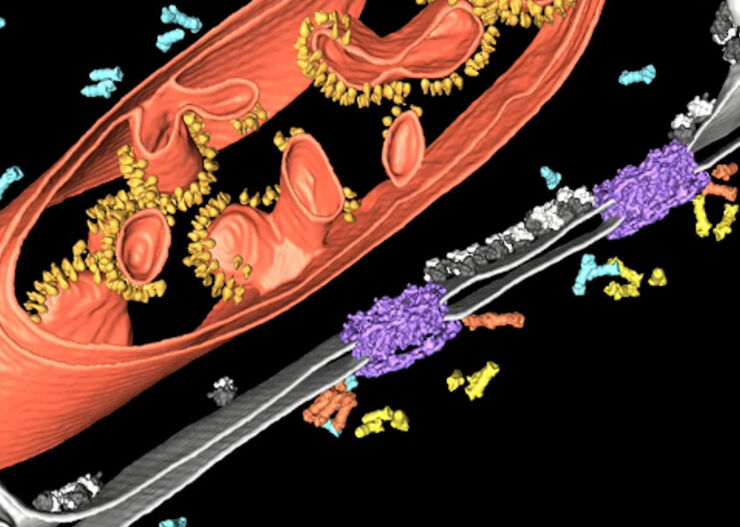
A Guide to Cryo-Electron Tomography
Cryo-electron tomography (CryoET) is used to resolve biomolecules within their cellular environment down to an unprecedented resolution below one nanometer.

The Cryo-CLEM Journey
This article describes the Cryo-CLEM technology and the benefits it can provide for scientists. Additionally, some scientific publications are highlighted.
Recent developments in cryo electron…

Targeting Active Recycling Nuclear Pore Complexes using Cryo Confocal Microscopy
In this article, how cryo light microscopy and, in particular cryo confocal microscopy, is used to improve the reliability of cryo EM workflows is described. The quality of the EM grids and samples is…
Improve Cryo Electron Tomography Workflow
Leica Microsystems and Thermo Fisher Scientific have collaborated to create a fully integrated cryo-tomography workflow that responds to these research needs: Reveal cellular mechanisms at…
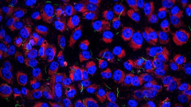
Imaging of Host Cell-bacteria Interactions using Correlative Microscopy under Cryo-conditions
Pathogenic bacteria have developed intriguing strategies to establish and promote infections in their respective hosts. Most bacterial pathogens initiate infectious diseases by adhering to host cells…
Fields of Application
Correlative Light and Electron Microscopy (CLEM)
Coral workflows by Leica Microsystems help users correlate fluorescence and electron microscopy (CLEM) data.
