Not all products or services are approved or offered in every market, and approved labelling and instructions may vary between countries. Please contact your local representative for further information.
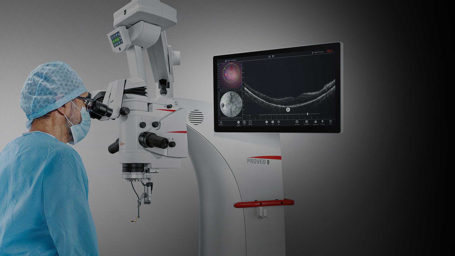
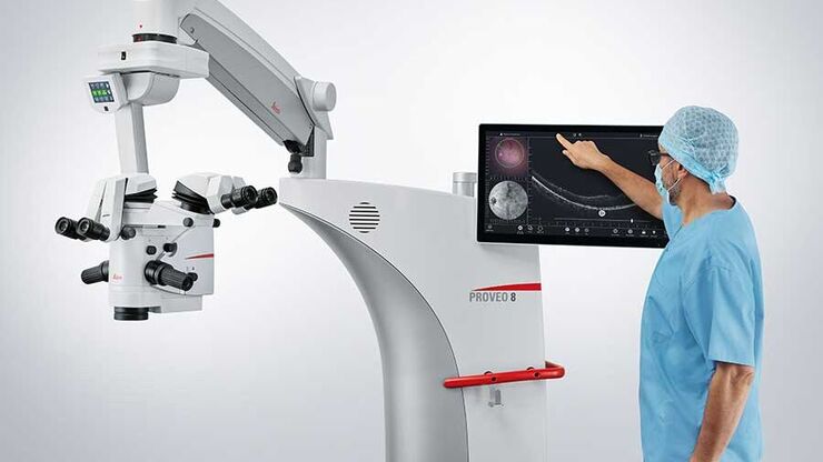
Apply your skills with even greater confidence during complex eye surgeries with a Leica OCT microscope solution.
EnFocus intraoperative Optical Coherence Tomography (OCT) allows you to see what lies underneath the surface. It gives you the additional real-time information you need for a deeper understanding of how subsurface tissue reacts to your surgical maneuvers.
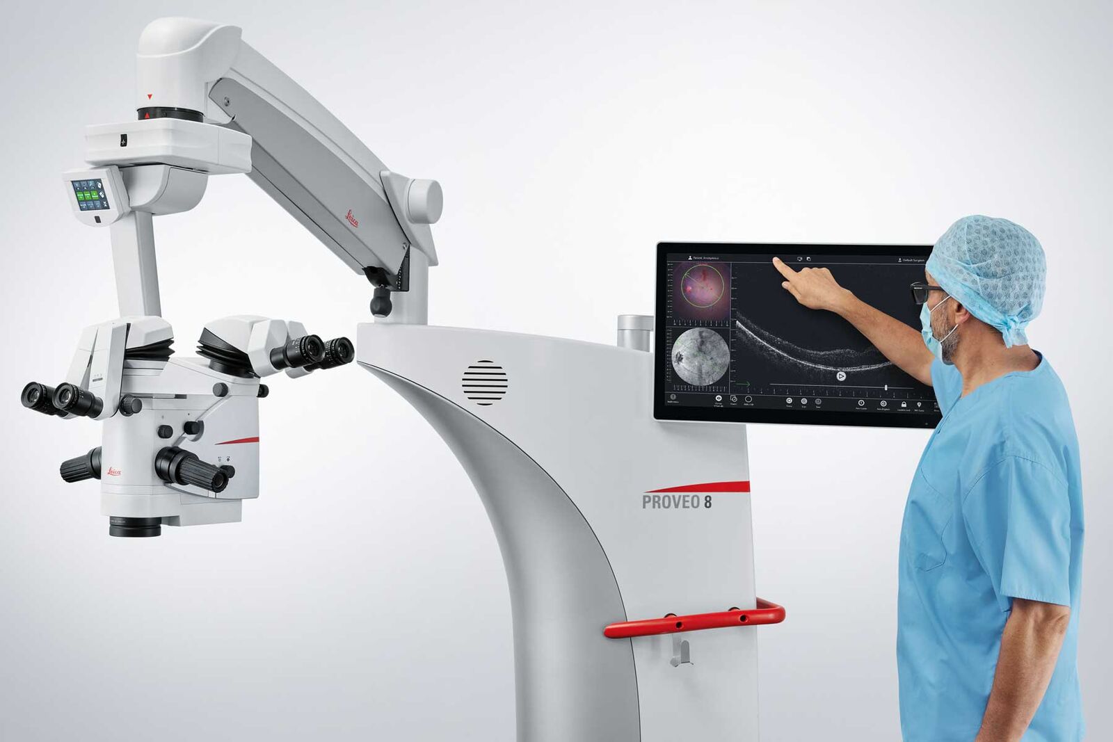
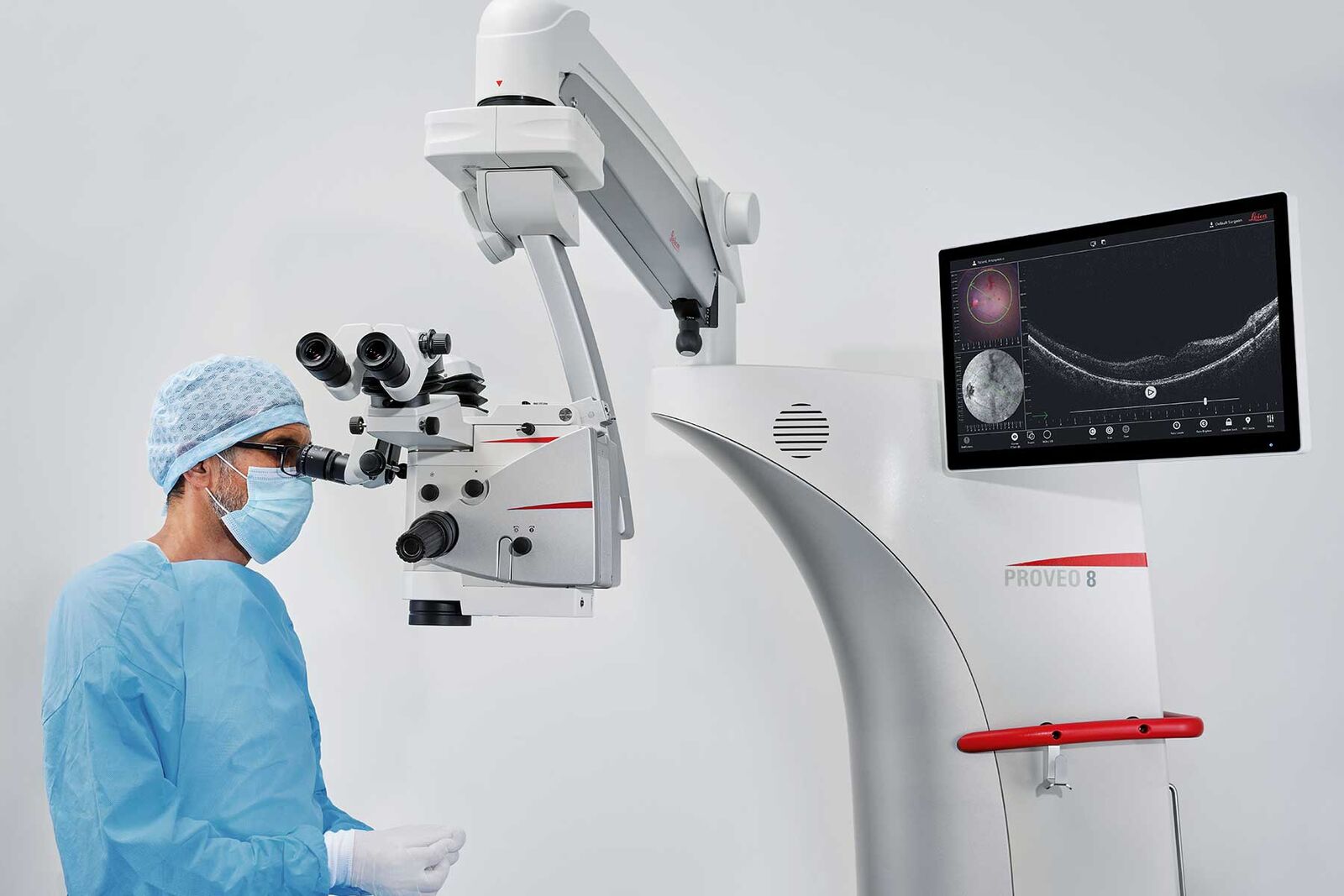
The EnFocus OCT imaging system is now also visually in perfect visual symbiosis with the Proveo 8 microscope. The optimized OCT scan head fits seamlessly into the optics carrier and allows for a more comfortable posture at the microscope.*
*Compared to the previous generation of EnFocus intraoperative OCT for the Proveo 8 floor stand microscope.
Not all products or services are approved or offered in every market and approved labeling and instructions may vary between countries. Please contact your local Leica representative for details.
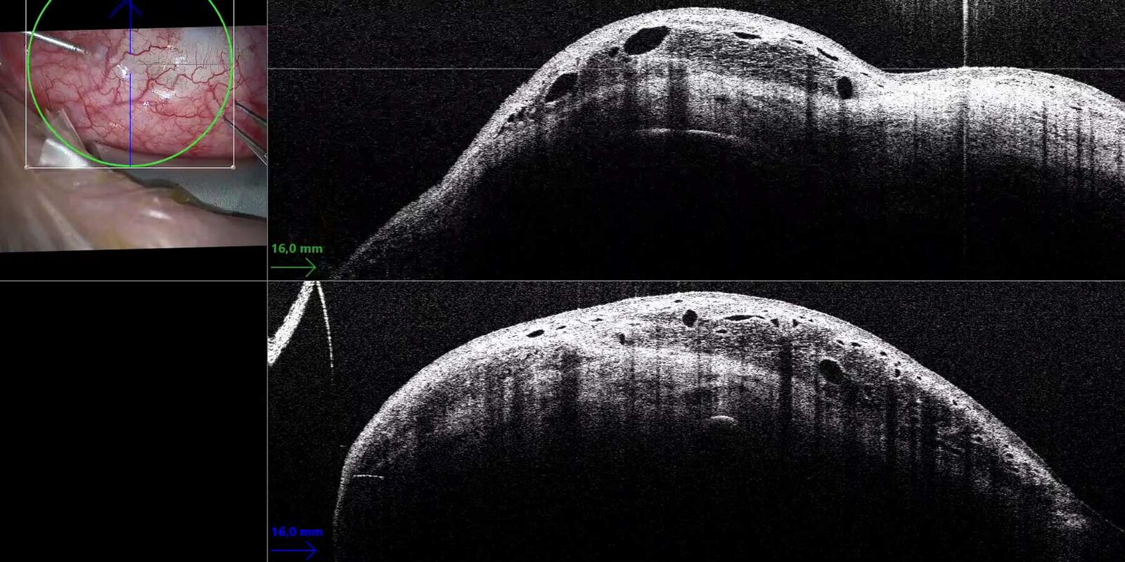
EnFocus offers wide scans, that cover the whole cornea and the anterior chamber – detail and resolution are impressive and allow a thorough visualization of anterior segment details. EnFocus provides a remarkable insight into the third dimension, meaning the vertical axis Z.
Greater insight: See more details below the surface
Supplement your microscope view with bright, sharp imaging of previously hidden subsurface tissue details. Intraoperative OCT imaging provides you with additional information on ocular pathology during surgery.
Clearly differentiate between artifacts and tissue due to the unique spectrometer technology with dispersion compensation software and a highly sensitive detector that captures more signal
See fine details even through blood in trauma cases thanks to an axial resolution of up to 4 μm in tissue
Capture comprehensive area scans with high lateral resolution, due to a high scan density of up to 1000 A-scans x 1000 B-scans to not miss out on an important detail
Playback acquired OCT scans via the microscope handle to check frame by frame or in video mode if, for example, sub-retinal fluid has remained, if the glaucoma drainage is positioned correctly or if the corneal graft is well opposed to the host cornea
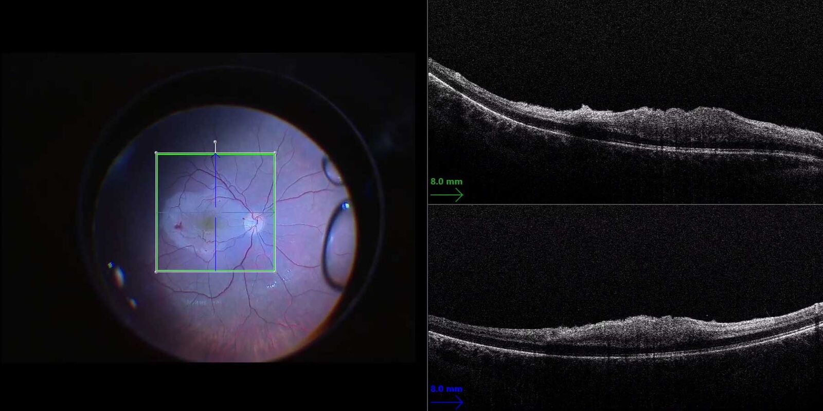
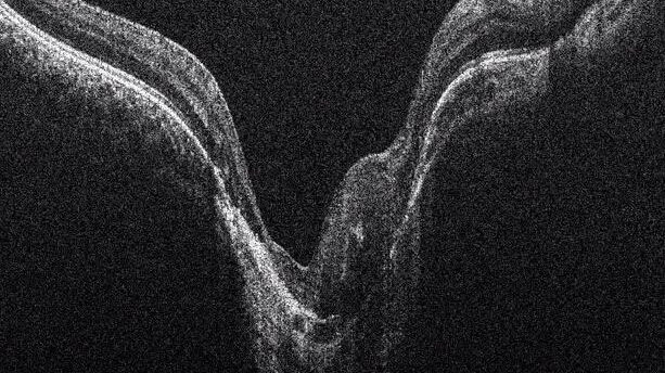
InVivo Software of the EnFocus intraoperative Optical Coherence Tomography (OCT) imaging system showing a cornea procedure in quadview. See the microscope image on the top left and the EnFace view on the bottom left, as well as the Optical Coherence Tomography (OCT) B-scan image on the right.
Greater insight: Get a wider view
Ensure you have a full view of the surgical field so you don’t miss features at the periphery. EnFocus provides a 20 x 20 mm lateral field of view even at high magnification.
No need to adjust the viewing perspective during surgery. This enables you to monitor any changes at the periphery even when working in the center of the eye.
Courtesy of University Hospital Dusseldorf, Germany imaged with Proveo 8 and EnFocus
Benefits for your surgery
Benefits for your retina surgery
Show details
Benefits for your retina surgery
Use OCT to assess the level of tension in a membrane peel in real time, in order to avoid potential tears and protect the integrity of underlying tissue. A high-resolution view of retinal membranes also aids examination of retinal morphology for residual membranes and complications such as macular holes, or sub-retinal edema.
Additional benefits for posterior surgery include:
Dynamic scan control via footswitch for swift adjustment of the scan angle to align with the membrane tissue
Easy integration of fundus viewing systems such as the BIOM5 from OCULUS with synchronized focus
Microscope view of the retina (left) supplemented with EnFocus OCT (right) to visualize membrane layers during membrane peeling. Image courtesy of Dr. Massimo D’Atri, Cagliari, Italy.
Benefits for your cornea surgery
Show details
Benefits for your cornea surgery
In advanced lamellar corneal surgeries such as DMEK (Descemet's Membrane Endothelial Keratoplasty) and DSAEK (Descemet’s Stripping Automated Endothelial Keratoplasty), this aids the surgeon in confirming the correct orientation of donor tissue.
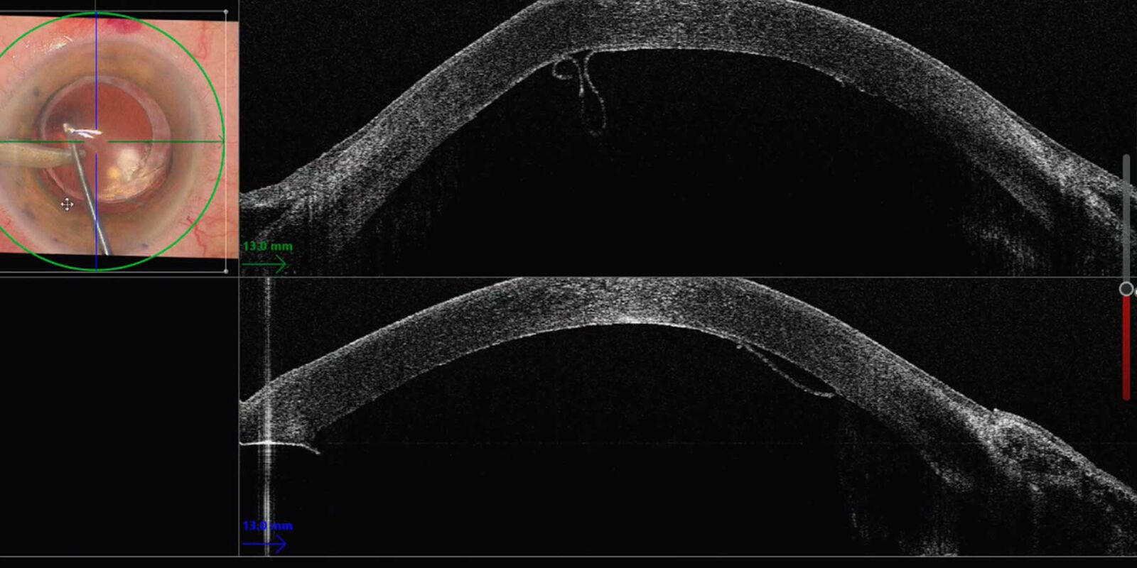
Thin Manual Descemet Stripping Endothelial Keratoplasty (TM-DSEK): The intraoperative OCT clearly showed the Descemet membrane peeling and endothelium. Image courtesy of Mr. David Anderson, MB BS FRCOphth PhD FEBO, University Hospital Southampton NHS FT, UK
Benefits for your glaucoma surgery
Show details
Benefits for your glaucoma surgery
OCT scans of up to 20 mm width support your view to aid e.g, accurate placement of an XEN gel stent in MIGS.
OCT can also support trabeculectomy by showing the needle positioning during the drug injection.
Intraoperative OCT picture showing 5-Fluorouracil (5-FU) drug injection after trabeculectomy. Images courtesy of Prof. Gerd Geerling, MD, PhD, FEBO, Department of Ophthalmology, University Hospital Düsseldorf, Germany
Immediate confirmation: Observe & respond to tissue changes
The EnFocus intraoperative OCT imaging system provides real-time confirmation of how ocular tissue is reacting to your surgical maneuvers. You can immediately adjust your surgical plan if needed for greater confidence in the surgical outcome.
Observe real-time tissue reaction and respond instantly
Real-time display of 30 fps provides immediate feedback at each step e.g. to verify adherence of donor tissue in DMEK or DSAEK surgery
If OCT reveals a complication which wasn’t visible via the microscope view, for example due to bleeding, you can instantly adapt your surgical plan
For additional verification you can easily review or playback through the acquired scans frame by frame or in video mode
Live on-screen measurements provide even more information e.g. cornea thickness and needle depth during DALK surgeries
Measurements with EnFocus intraoperative OCT during deep anterior lamellar Keratoplasty for Keratoconus (DALK) help to quantify the incision depth. Courtesy of Enrico Bertelli MD,Head of the Ophthalmic Dept.,Bolzano Hospital, Italy
EnFocus built into the Proveo 8 supports your workflow and enables you to independently control your OCT view. You are free to concentrate on your procedure.
Easily activate intraoperative OCT yourself at any point during surgery via footswitch, handle, or 27” touch screen HD monitor
Preprogram your personal settings and modes before surgery according to surgery type and step then allocate to the footswitch or handle for a smooth workflow
Record or capture images in the same way with the Evolution4K recording system from Med X Change for comprehensive documentation
View your microscope and intraoperative OCT-image on the 27” HD touch screen monitor and for even larger screen projections, four video outputs are available on the Proveo 8
Assistant initiating recording via touch screen.
Optimized design for enhanced work comfort
The height-reduced, slimmed-down new scan head of the EnFocus OCT system fits seamlessly into the design of the Proveo 8 optics carrier. You benefit from more comfort as you can adopt a more relaxed posture at the microscope, due to the reduced height of the optics carrier with the OCT attachment. *
Your OR staff will also feel the difference, as there is more free space behind the optics carrier, which can simplify handling of the sterile field.
*Compared to the previous generation of EnFocus intraoperative OCT for Proveo 8 floor stand microscope
Side view of the optimized design of the EnFocus OCT scan head, available for the Proveo 8 ophthalmic microscope in the floor stand configuration.
Workflow freedom in the operating room
Hear from vitreoretinal surgeon Dr. Barbara Parolini, Eyecare Clinic Brescia, Italy, about surgical freedom with the EnFocus OCT imaging system, thanks to the almost full self-operation during surgery.
Dr. Barbara Parolini, Eyecare Clinic Brescia, Italy
Maximum freedom: OCT imaging system, with intuitive control
No need to interrupt surgery or rely on a technician to acquire an optimal OCT image thanks to automated functions and intuitive operation.
You can activate automatic image optimization with just a tap of the footswitch, handle or touch screen. Brightness and sharpness of the image is adjusted for you, so you can rest assured that you always have an optimal OCT image. The Auto Locate function centers the OCT image automatically. Location lock in the z-direction then ensures the image remains centered throughout your procedure. It's just as easy to adjust scan pattern, scan size and scan density.
See how footswitch control offers maximum freedom in operating the EnFocus intraoperative OCT system
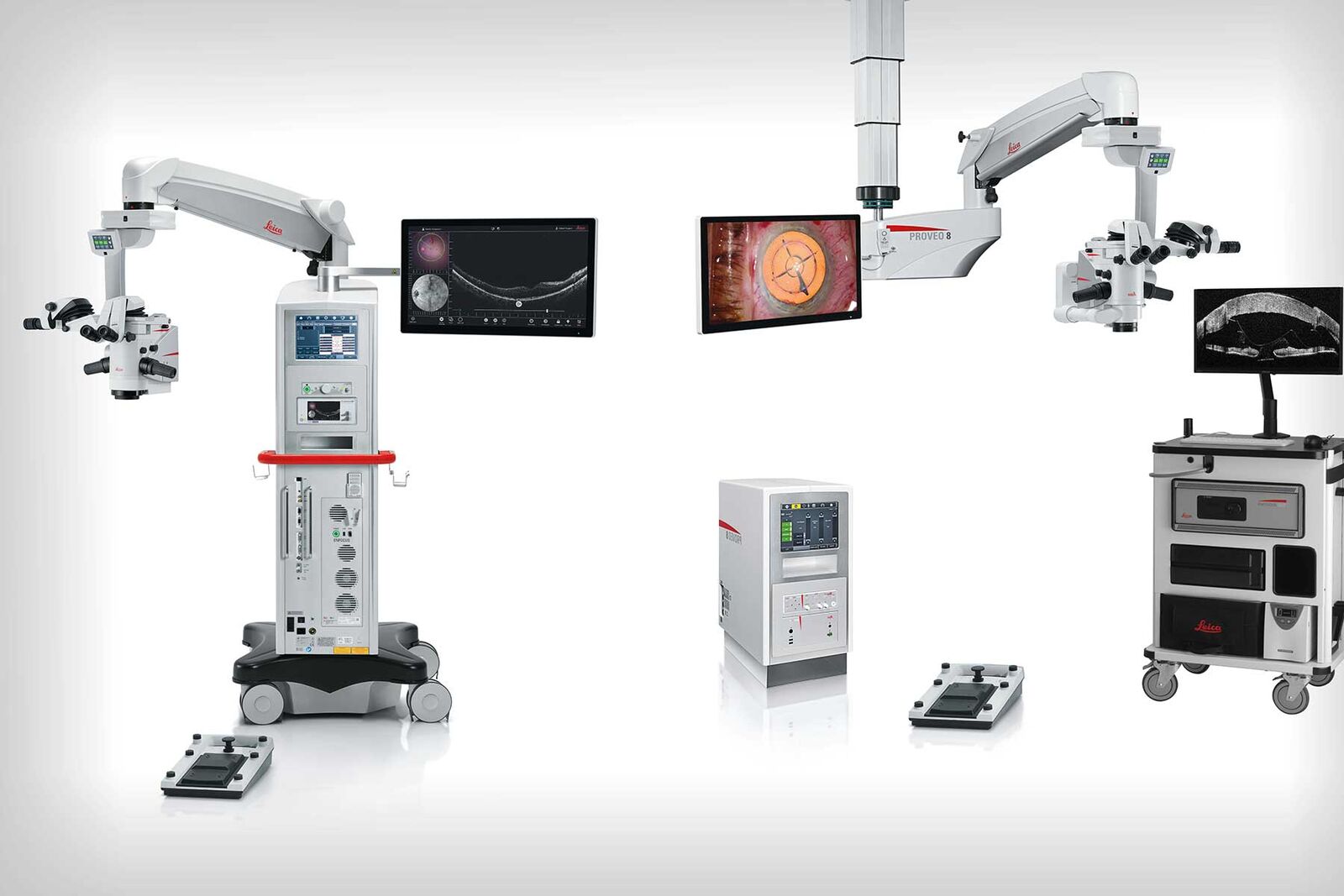
One OCT imaging system for all your needs
The EnFocus intraoperative OCT imaging system for the Proveo 8 ophthalmic microscope offers you an OCT microscope solution that precisely fits your needs. Whether for anterior or posterior surgery, for use in a large OR or a small surgery center, there is a solution to meet your requirements:
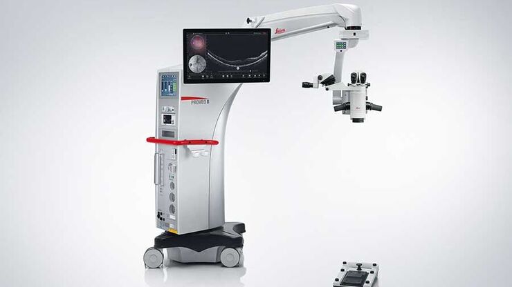
Proveo 8 floor stand with built-in EnFocus OCT
Proveo 8 telescope ceiling mount with EnFocus OCT (external unit)
Left: Proveo 8 floor stand microscope with built-in EnFocus intraoperative OCT.
Right: Proveo 8 telescope mount CT42 with external tower unit & EnFocus intraoperative OCT cart solution (standard scan head design).
The information provided on this page is intended for healthcare professionals. Please note that this information is not intended for the general public.