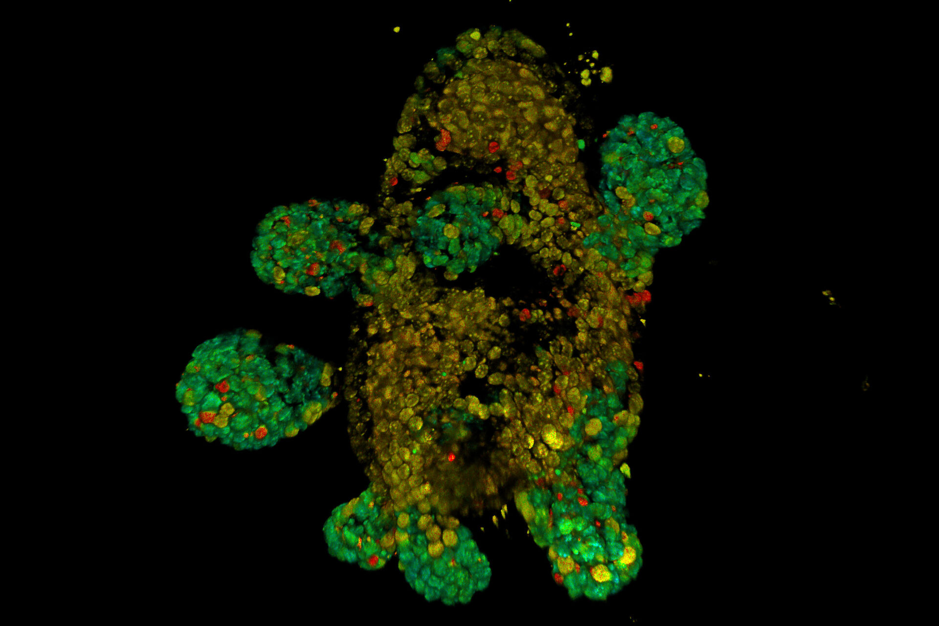
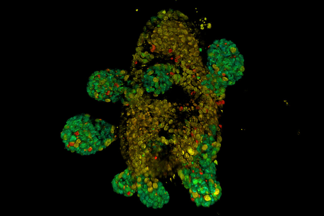
Cancer often comes with changes in the metabolism of cells. Monitoring the metabolic state using fluorescence lifetime imaging microscopy (FLIM) is a good way to assess these changes. In some cases, endogenous signals, e.g., from NAD/NADH, can be exploited, while in other cases the resulting information is not sufficient. In this publication, the mitochondrial membrane potential (MMP) was studied using different fluorescent probes (SYTO dyes and TMRM) in various samples, including organoids derived from mouse intestine. The authors showed that the dyes could quickly and efficiently stain the organoids and that the stained organoids could be imaged using two-photon excitation. With this approach, the authors could identify the active population of cells in the stem cell niche. Results for this study were obtained using a Leica multiphoton microscope with FLIM. For more details, refer to the publication.


 in 14 x 18 tiles. Lifetime gives an additional contrast that allows to differentiate different structures in histological stainings.")
