Robert Ranner

Robert Ranner, Product Manager EM Specimen Preparation at Leica Microsystems, Nanotechnology Division. He is responsible for solid state preparation instruments. Robert has ten years application experience in industrial sample preparation for TEM, SEM.
 chip cross section acquired at higher magnification showing a region of interest.")
Structural and Chemical Analysis of IC-Chip Cross Sections
This article shows how electronic IC-chip cross sections can be efficiently and reliably prepared and then analyzed, both visually and chemically at the microscale, with the EM TXP and DM6 M LIBS…

High Resolution Array Tomography with Automated Serial Sectioning
The optimization of high resolution, 3-dimensional (3D), sub-cellular structure analysis with array tomography using an automated serial sectioning solution, achieving a high section density on the…
Practical Applications of Broad Ion Beam Milling
Mechanical polishing can be time consuming and frustrating. It can also introduce unwanted artifacts when preparing cross-sectioned samples for electron backscatter diffraction (EBSD) in the scanning…
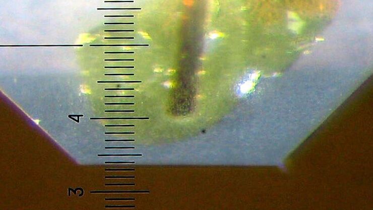
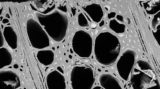
Brief Introduction to Specimen Trimming
Before ultrathin sectioning a sample with an ultramicrotome it has to be pre-prepared. For this pre-preparation, special attention must be paid to the sample size (size of the section), location of…
