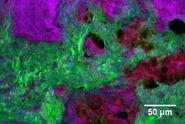
, unsaturated lipids (magenta, 3050 cm-1), collagen (SHG, cyan). Sample courtesy of R. Rudolf, J Klicks, Hochschule Mannheim")
A true multi-modal optical discovery platform
The STELLARIS 8 CRS coherent Raman scattering microscope offers the two popular CRS modalities – Stimulated Raman Scattering (SRS) and Coherent anti-Stokes Raman Scattering (CARS) – and allows for the simultaneous acquisition of two-photon fluorescence and second-harmonic generation signals. The seamless integration of CRS with the STELLARIS confocal fluorescence microscopy platform results in a true multi-modal optical discovery platform that is capable of capturing a unique combination of biochemical, biophysical, and molecular contrasts.
5-minute overview of the benefits of label-free chemical imaging with the STELLARIS 8 CRS for life science research
5-minutes of background information on the CRS technique and the hands-free standalone STELLARIS 8 CRS solution
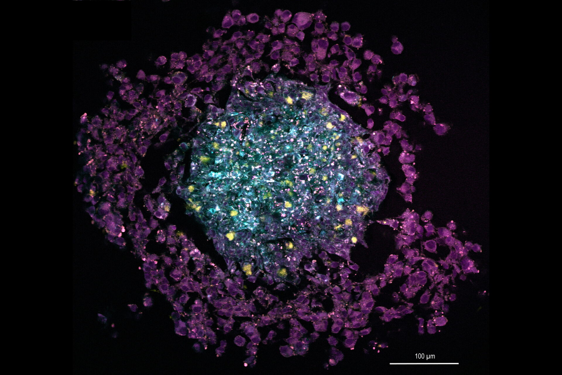
Multi-color SRS image of a tri-cellular cancer spheroid

 dataset, showing the biochemically distinct structures of a fresh, untreated apple slice.")