

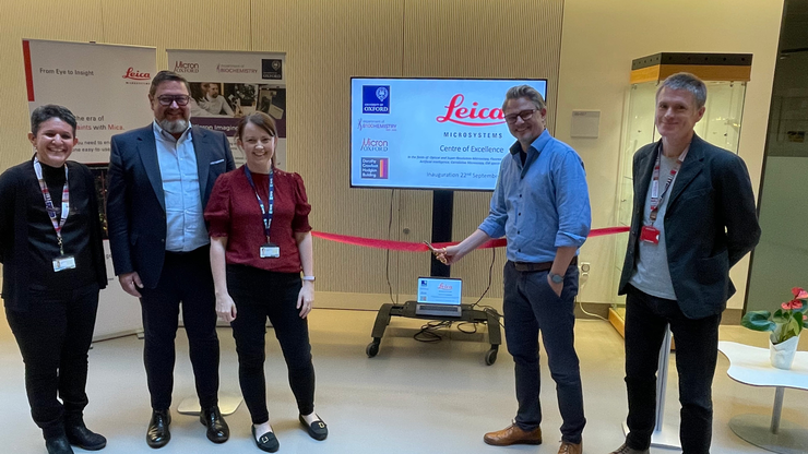
Leica Microsystems, together with the Centre of Excellence at the Micron Bioimaging Facility within the Department of Biochemistry, University of…
Centre of Excellence at the Micron Bioimaging Facility
Established in 2023, the Centre of Excellence welcomes researchers from around the world, giving access to Leica Microsystems’ state-of-the-art optical microscopy systems for routine and advanced (sub)cellular imaging. Located within the Department of Biochemistry, the staff at the Micron Bioimaging facility provide world-class education and training to students and professionals, helping scientists to push the boundaries of understanding of the inner workings of cells and tissues.
Uniting the microscopy expertise of Leica Microsystems with Oxford's academic excellence, the mission of the Centre of Excellence is to advance scientific research and champion state-of-the-art techniques in optical and super-resolution microscopy, artificial intelligence (AI), correlative microscopy, and electron microscopy (EM) specimen preparation.
Find out more
Visit the Micron Bioimaging Facility website to see how you can access advanced imaging technology, with expert guidance and support for your project.
Latest Updates
Super-resolution microscopy system at the heart of groundbreaking research recognized by Cancer Research UK’s Research is Beautiful campaign
Leica Microsystems has announced an exciting new collaboration with the Department of Biochemistry at The University of Oxford UK, in the fields of…
Empowering the scientific community through shared knowledge
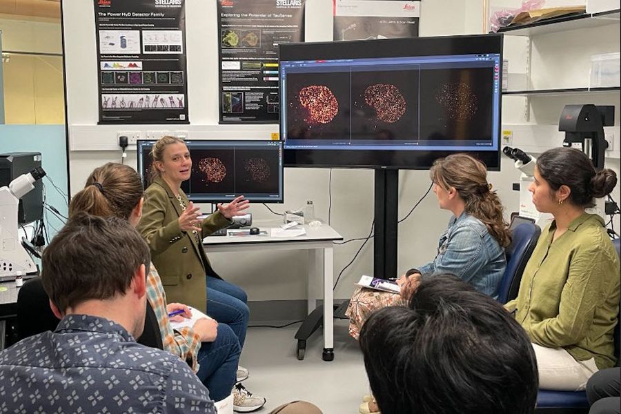
The centre fosters a collaborative training culture that advances learning and nurtures future talent. Experts from across the scientific community can meet, exchange insights, and expand their expertise through workshops and seminars.
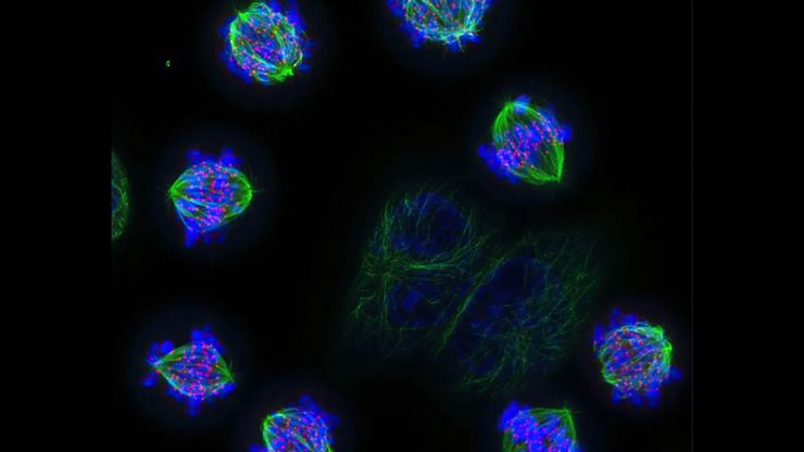
Within the facility, researchers gain access to the latest advanced confocal and widefield microscopy systems and benefit from immersive training sessions conducted by Leica Application Specialists. Scientists can harness microscopy workflows and technologies such as super-resolution live-cell imaging with the latest generation STELLARIS confocal microscope systems featuring TauSTED Xtend. The facility also houses a THUNDER Imager Cell, which is heavily used by researchers for a variety of widefield imaging applications. They can also take advantage of cryogenic fluorescence imaging for correlation with cryo-electron microscopy (cryoCLEM), in collaboration with the neighbouring COSMIC CryoEM facility.
Additionally, the facility offers a dedicated analysis suite equipped with Aivia AI Image Analysis Software.
Imaging in action at Micron
From cellular dynamics to complex tissue analysis, the researchers that Micron supports are using Leica microscopy systems to investigate biological processes and address key scientific questions in life science research.
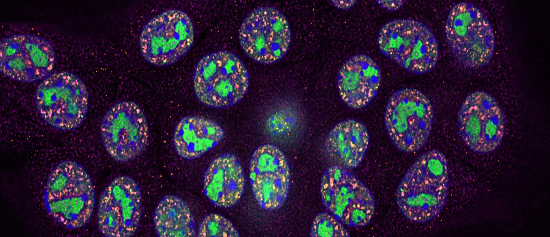
Investigating pancreatic ductal adenocarcinoma
Dr. Zeinab Rekad from the Department of Biochemistry is interested in whether RNA transcription and maturation are globally dysregulated under conditions that mimic the early stages of Pancreatic Ductal Adenocarcinoma. Immortalised mouse pancreatic cells were exposed to a uridine analogue, which is incorporated into newly transcribed RNA. This was followed by click chemistry. To detect and count the number of nucleoli and RNA splicing regulatory units (speckle units) per cell, Dr. Rekad used the THUNDER Imager Cell within the Micron Bioimaging Facility. By applying the THUNDER Small Volume Computational Clearing (SVCC) method, she was able to remove out-of-focus blur and generate high resolution, high contrast images. The signal intensity of the nucleoli was used to assess the level of rRNA expression. Finally, the shape of the structures was assessed using masks and shape descriptors analysis.
Understanding molecular mechanisms in the nervous and vascular systems
Prof. Elena Seiradake, also within the Department of Biochemistry is interested in the mechanisms that guide cells to form complex tissues such as neural and vascular networks. Her group has been using the STELLARIS system at Micron to get some very exciting results.


Incorporating the Leica STELLARIS into our research has significantly advanced our ability to directly observe distinct molecular assemblies that instruct cell behaviour. The nanoscale resolution provided by STED microscopy has opened new dimensions in our understanding of the fundamental mechanisms by which receptors control biological processes in neural and vascular systems.
This partnership provides users with direct access to Leica Microsystems’ cutting-edge microscopy technologies and global expertise. Together, we’ve built an environment where researchers can tailor their imaging workflows and engage closely with both Leica and Micron to advance their scientific goals.
Apply now
Register your interest in becoming a facility user.
Micron, Oxford related articles

Unlocking the Secrets of Organoid Models in Biomedical Research
Get ready to delve deeper into the world of organoids and 3D models, which are essential tools for advancing our understanding of human health. Navigating these complex structures and obtaining clear…

Revealing Neuronal Migration’s Molecular Secrets
Different approaches can be used to investigate neuronal migration to their niche in the developing brain. In this webinar, experts from The University of Oxford present the microscopy tools and…
Meet the Team at the Micron Bioimaging Facility

Lothar Schermelleh, Professor of Bioimaging & Academic Director
Lothar joined the Department of Biochemistry in 2011 as Micron Senior Research Fellow and Principal Investigator, where he led the development of computational analysis tools for standardised quality control and improved fluorescence labelling protocols for super-resolution 3D structured illumination microscopy (3D-SIM). His biological research aims to understand the relationship between 3D nuclear organisation, chromatin structure and genome function in mammalian cells, using advanced optical imaging. The lab currently focuses on uncovering the regulatory role and interplay between biophysical forces, epigenetic memory, and cohesin complex activity to modulate cell-type-specific transcriptional programmes. In November 2020 he became Academic Director of Micron. In the same year he was named Associate Professor and then made Professor in 2024.

Dr. Deirdre Kavanagh, Manager, Micron Bioimaging Facility
In May 2021, Deirdre joined the Department of Biochemistry as the Facility Manager for Micron. An interdisciplinary scientist specialising in super-resolution microscopy, fluorescence correlation spectroscopy, and light-sheet technologies, she holds a PhD in Engineering and Physical Sciences. Deirdre is primarily responsible for overseeing the day-to-day operations of the Micron facility. During her postdoctoral research, she applied advanced super-resolution microscopy to study the protein interactions, organisation, and dynamics that govern cell communication. In addition to managing the facility, she is actively involved in organising and developing microscopy workshops, training courses, and public engagement initiatives. She serves on several microscopy committees, including the Royal Microscopical Society Light Microscopy Committee.

Dr. Niloufer Irani, Deputy Manager, Micron Bioimaging Facility
Niloufer joined Micron in 2021 from the Department of Plant Sciences, University of Oxford with research interests in the molecular mechanistics of endomembrane organisation, transport, and its links to signalling. Taking up the opportunity to work at the multi-million-pound facility at Micron, she brings out-of-the box thinking to experimental questions that are considered throughout the world.. An independent scientist, she has studied and worked globally in India, the USA, Belgium and the UK. Niloufer is involved in all aspects of management of the facility, teaching, training, consulting, sample preparation and maintenance of the instruments. She actively organises Leica workshops with the Leica application specialists to share microscopy best practice to all users.
