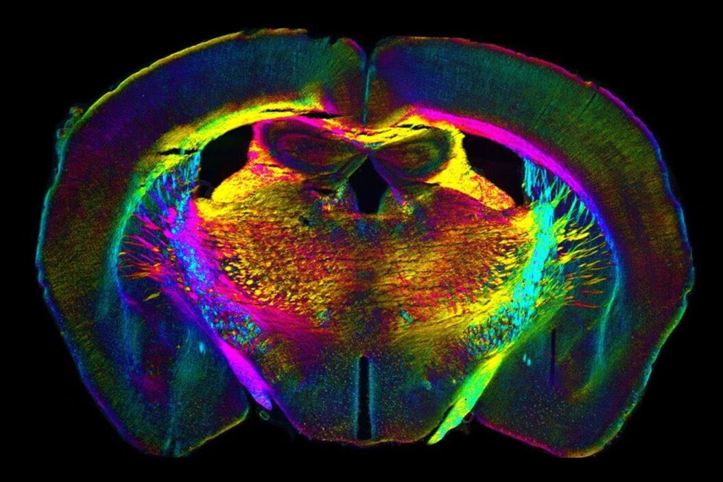
In the last 3 years, microscopists have started to use "AI based" solutions for a wide range of applications, including image acquisition optimization (smart microscopy), object classification, image classification, segmentation, restoration, super resolution and virtual staining.
The 6th edition of the AI Microscopy Symposium offered a unique forum for presenting and discussing the latest AI-based technologies and tools in the field of microscopy and biomedical imaging. Additionally, the symposium highlighted scientific breakthroughs which are enabled by machine learning or deep learning technologies.


