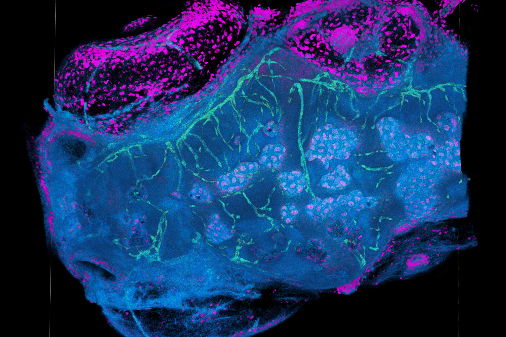
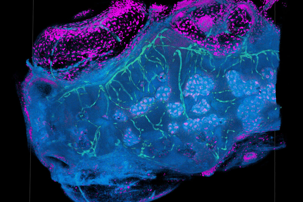
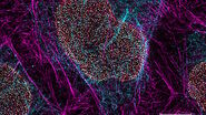
Read the Application Note to find out how you can use TauSense technology to:
- Explore a new dimension of information in every confocal experiment with instant access to functional information.
- Improve image quality by removing unwanted fluorescence signals that mask your data and prevent you from truly seeing your sample.
- Separate fluorescent species based on lifetime information, allowing you to distinguish fluorescent signals with similar spectra.
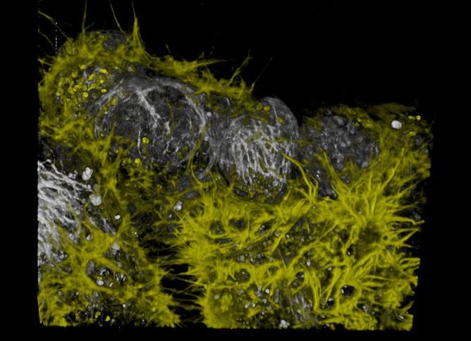
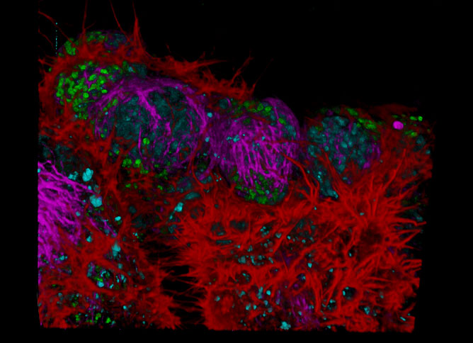
Fluorescence lifetime-based multicolor imaging in live NE-115 cells. Actin: LifeAct-mNeonGreen (left: yellow, right: red); mitochondria: MitoTracker Green (left: yellow, right: green); nuclei: NUC Red (left: gray, right: blue); and tubulin: SiR-tubulin (l
