Super-Resolution imaging with STELLARIS confocal platform
The STELLARIS confocal platform gives access to super-resolution with LIGHTNING and STED technologies for visualizing structures with greater precision and uncovering details that would otherwise not be visible.
TauSTED Xtend delivers details at the nanoscale level on-the-fly
Supplementary movie 1 from application note “Gentle, Multicolor Live imaging at the Nanoscale with TauSTED Xtend". Download the application note now.
Gentle multicolor live cell TauSTED Xtend 775
Supplementary movie 2 from application note “Gentle, Multicolor Live imaging at the Nanoscale with TauSTED Xtend". Download the application note now.
Long term TauSTED Xtend on green fluorescent proteins
Supplementary movie 3 from application note “Gentle, Multicolor Live imaging at the Nanoscale with TauSTED Xtend". Download the application note now.
Two-color TauSTED Xtend 589 of the inner ear section of a mouse

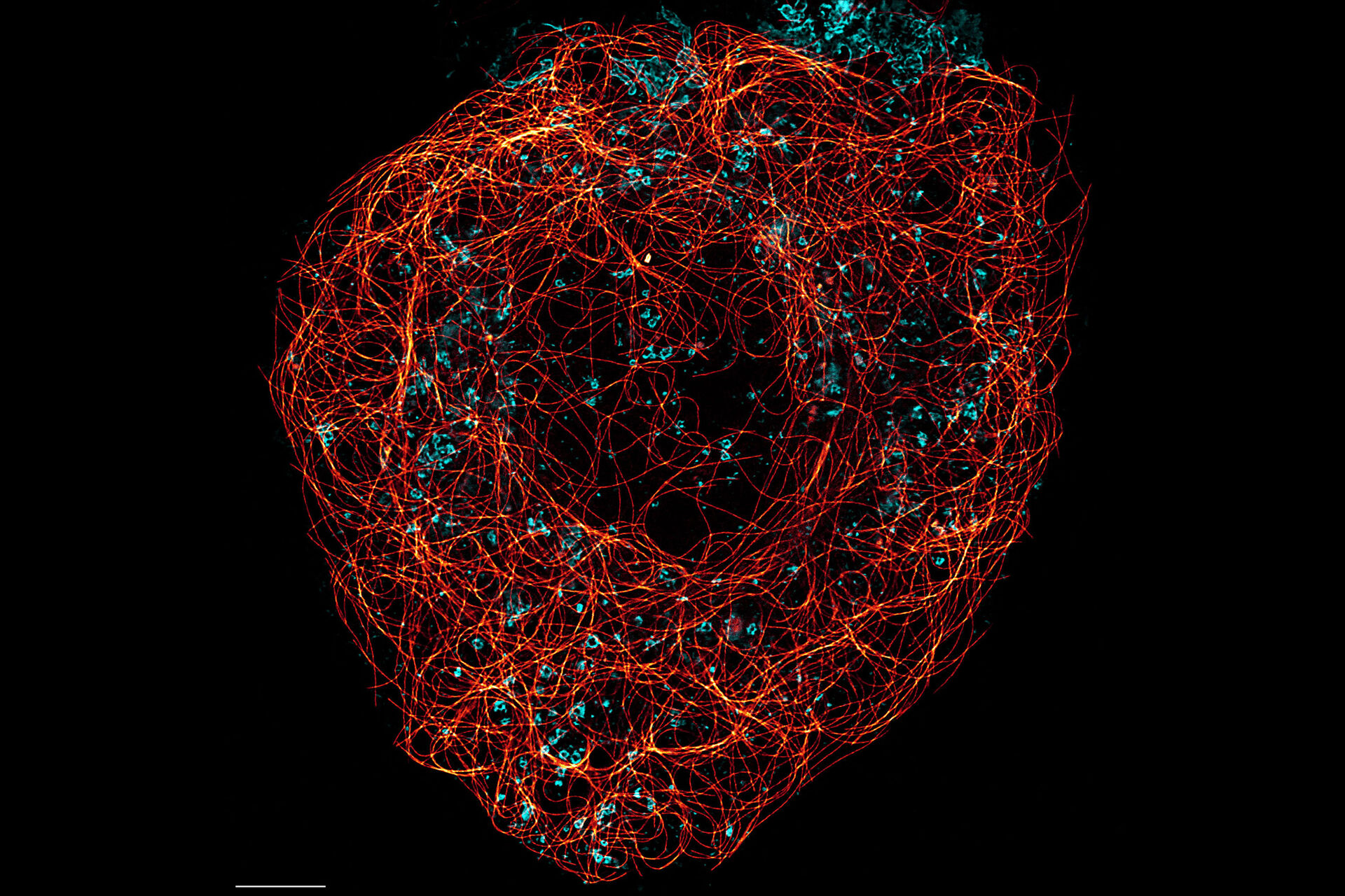
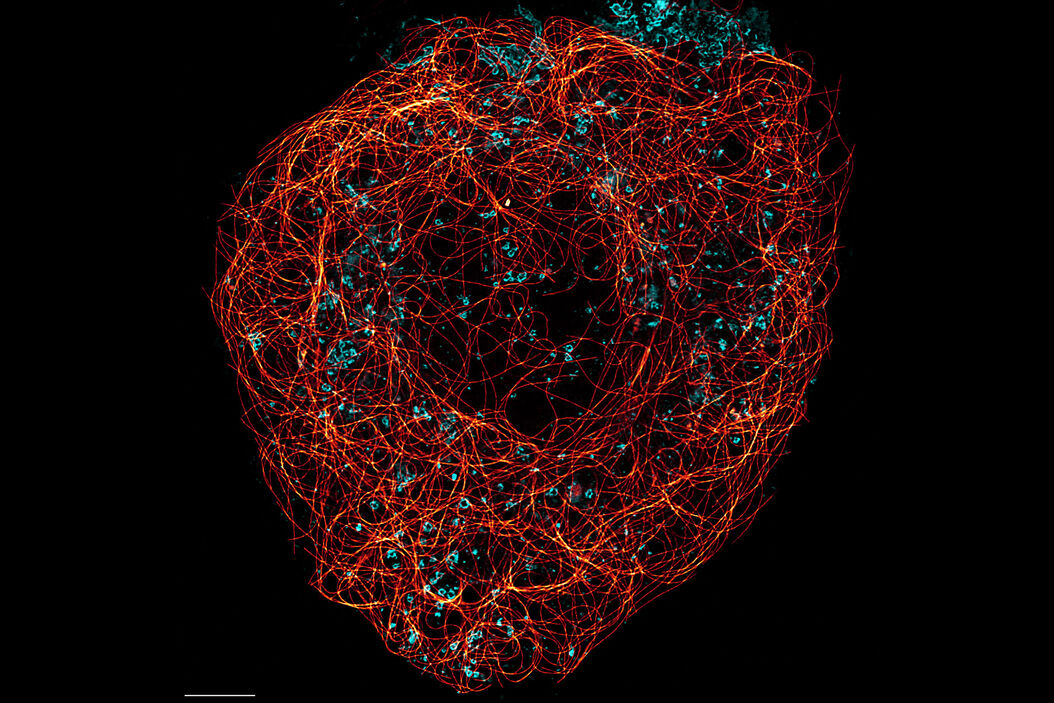
Cytoskeleton and membranes in live cell imaged with TauSTED
Image acquired with STELLARIS STED.


TauSTED 775 resolves the intricate cytoskeleton network labeled with SiR-tubulin (glow - Spyrochrome), and trafficking vesicles labeled with CF594 (cyan - Biotium).
Cytoskeleton and membranes in live cell imaged with TauSTED
Image acquired with STELLARIS STED. SiR and SPY are available from Spirochrome. CF dyes are available from Biotium Inc.


Live cell TauSTED on U2OS cells, using labels for actin (SiR-actin, glow), microtubules (SPY555-tubulin, cyan), and membranes (CF488A coupled to WGA, green). Scale bar: 10
STED for malaria research

Three color STED imaging of Cos 7 cells

3D STED 775 deep nanoscopy of glomerulus in cleared kidney tissue

STED for Microbiology
Image acquired with STELLARIS confocal platform.


, actin network (ATTO 647N), and nuclear pore basket (CF 680R).")