Tissue imaging with STELLARIS confocal platform
The STELLARIS confocal platform gives you access to multiphoton imaging, STED super-resolution, fluorescence lifetime imaging microscopy (FLIM), and Digital Light Sheet (DLS), to image the most demanding tissue samples. This gallery shows images from tissue obtained with the STELLARIS confocal platform.
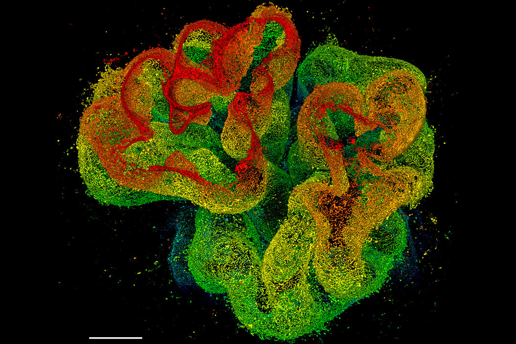
3D STED 775 deep nanoscopy of glomerulus in cleared kidney tissue

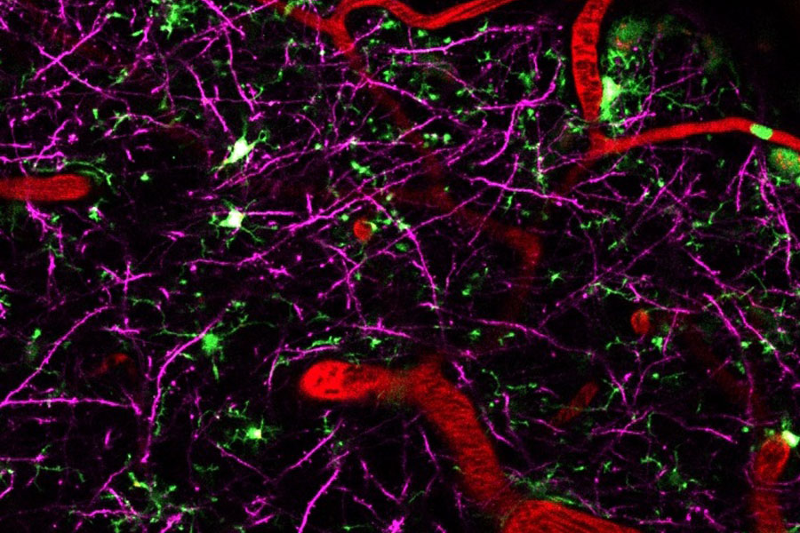
4 colour live imaging of mouse brain cortex
Neurodegenerative diseases are usually caused by a combination of different factors. Being able to visualize multiple players at once greatly helps to investigate and understand how different compoonents interact and influence each other. To do this, a microscope that combines deep tissue penetration with spectral flexibility to enable multicolor imaging liek STELLARIS 8 DIVE is the best choice.

Multicolor Deep In Vivo Imaging to uncover the interplay of various players in neurodegenerative diseases
Neurodegenerative diseases are usually caused by a combination of different factors. Being able to visualize multiple players at once greatly helps to investigate and understand how different compoonents interact and influence each other. To do this, microscope that combines deep tissue penetration with spectral flexibility to enable multicolor imaging like STELLARIS 8 DIVE is the best choice.
Expand the potential of deep in vivo experiments with label-free imaging
Molecules such as collagen and elastin have relevant roles in diseases like cancer. Our 4-tune detector enables the use of second and third harmonic generation signals that allow you to study these important structures without staining. The combination of DIVE with STELLARIS also enables the use of lifetime-based information intrinsic to fluorescence. This ability allows you to perform experiments like metabolic mapping of a specimen via lifetime imaging of NADH or FAD.

Understand cancer better with deep in vivo imaging
Multiphoton systems are usually rigid to use and need to be adapted to each experiment and user. Add to this the stress of working with live animals or freshly explanted tissue and you quickly understand the advantage of having flexibility when doing multiphoton experiments. STELLARIS 8 DIVE provides you with an easy, hassle-free workflow from setup to final results thanks to the seamless integration of multiphoton capabilities into the STELLARIS software.

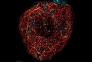

, actin network (ATTO 647N), and nuclear pore basket (CF 680R).")