What is phase contrast?
Phase contrast is an optical contrast technique for microscopy which makes unstained structures in the cells of biological specimens visible. Cell structures that appear transparent with brightfield illumination can be viewed in high contrast and rich detail using phase contrast. Differences in optical density between structures in the cell can cause light that interacts with them to attain a phase shift. This phenomenon is the basis of phase contrast. As a result, more optically dense structures will look darker than less optically dense ones.
Related articles
Optical Contrast Methods

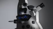
Phase Contrast and Microscopy
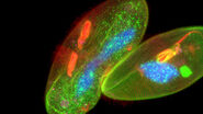
 cells taken with phase contrast.")
Introduction to Widefield Microscopy

What are some phase contrast microscope uses? Which kind of samples can you visualize?
Most often biological specimens and tissues are observed with a phase contrast microscope. A large variety of biological specimens can be observed with phase contrast from fixed specimens to living cells and tissues.
Life Science Research
Leica microscopes providing phase contrast are commonly used in life science research for the visualization, analysis, and documentation of biological structures and cellular processes.
Materials & Earth Science
Leica microscopes capable of phase contrast make a difference for the study of transparent and colorless minerals, crystals, and polymers.
Forensics
For forensic applications concerning the evidentiary investigation of paints, pigments, textiles, fibers, and human tissues, Leica microscopes offering phase contrast are very useful solutions.

How does a phase contrast microscope work?
A phase contrast microscope is similar to a conventional widefield microscope, except it uses an aperture in the shape of an annulus and a quarter-wave (λ/4) phase plate. The annular aperture is placed between the light source and condenser lens and the phase plate after the objective inside the microscope optics. Ring-shaped light that passes through the aperture is focused by the condenser onto the biological specimen to be observed.
Portions of the ring-shaped light are diffracted by optically dense structures of the specimen and experience a negative phase shift of about λ/4. This phase-shifted, diffracted light bypasses the λ/4 plate. In contrast, the portion of the ring-shaped light that passes directly through the specimen non-deviated will hit the phase plate which causes a positive λ/4 phase shift. As the total difference in phase shift between the light diffracted by the specimen’s structures and that which passes through phase plate will be about λ/2, destructive interference will occur. Consequently, more optically dense structures will appear darker than those that are less optically dense.


Using phase shifts for image formation
Related articles
Introduction to Mammalian Cell Culture

How to do a Proper Cell Culture Quick Check

How to Determine Cell Confluency with a Digital Microscope

Frequently asked questions phase contrast microscopes
The phase contrast method for microscopy was developed in the 1930s by the Dutch physicist Frits Zernike. After 1942, it became a widely used microscopy technique. In 1953, Zernike was awarded the Nobel Prize for Physics. For more details, refer to the articles: A Brief History of Light Microscopy – From the Medieval Reading Stone to Super-Resolution & Phase Contrast
A phase contrast microscope is similar to a conventional brightfield microscope, except it uses an annular aperture in front of the light source and a quarter-wave phase plate after the objective lens. For more information, refer to the article: Phase Contrast
Yes, a phase contrast microscope can be equipped with a camera for recording images observed with the contrast method.
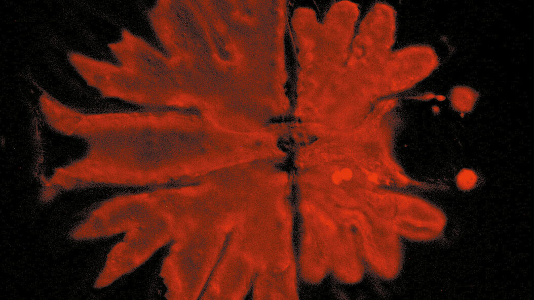
 cells taken with phase contrast.")