Contact Us

Live Cell Imaging

Leica Microsystems provides you with intelligent innovations in live-cell imaging. Our solutions help you get the best image quality while protecting your samples.

With the variety of fluorescent proteins and multicolored probes that have been developed, it is now possible to label virtually any molecule. The ability to visualize protein dynamics in vesicles, organelles, cells, and tissues has provided new insights into how cells function in healthy and disease states. These insights include the spatiotemporal dynamics of processes like mitosis, embryonic development, and cytoskeleton changes. When studying live cells, common obstacles include phototoxicity and photodamage. To capture fast biological processes, it is crucial to keep the cells healthy and obtain crisp images for reliable data that are free of artifacts. Live‐cell microscopy often requires a compromise between image quality and cell health. During imaging, certain environmental conditions must be maintained to avoid changes in the cells. 

A variety of high-performance Leica live cell imaging solutions can overcome these challenges for live-cell imaging enabling new information for cellular physiology and dynamics to be discovered. 

Simply get in touch!

Our experts on solutions for live cell imaging applications are happy to help you with their advice.

Your Live Cell Imaging Needs

To perform successful live-cell imaging experiments, using the right platform is critical. When choosing an optical microscope for live‐cell imaging, the following 3 variables should be considered: detector sensitivity (signal‐to‐noise ratio), specimen viability, and image-acquisition speed.

Methods suitable for live-cell applications enable visualization of the dynamics without causing cell damage, as it can affect the results.

Leica Microsystems offers the latest innovations in widefield and confocal imaging technologies for fast 3D live cell imaging with the THUNDER Imagers, Mica – the world’s first Microhub, STELLARIS confocal platform, and FLIM.

Live‐cell imaging is mainly performed with fluorescence microscopy. Widefield microscopy, providing flexible excitation and fast acquisition, is typically used to visualize cell dynamics and development over long times. Confocal microscopy is typically used to study subcellular dynamic events. Multiphoton microscopy allows excitation with longer wavelength light reducing photobleaching and extending cell viability. Finally, fluorescence lifetime imaging (FLIM) can be applied to study fast dynamic signaling events in cells.

Live Cell Imaging System DMi8

The modular DMi8 inverted microscope is the heart of the DMi8 S platform solution. For routine to live cell research, the DMi8 S platform is a complete solution. Whether you need to precisely follow the development of a single cell in a dish, screen through multiple assays, obtain single molecule resolution, or tease out behaviors of complex processes, a DMi8 S system will enable you to see more, see faster, and find the hidden.

Cell Viability & Dynamics During Live Cell Imaging

Leica Microsystems provides you with intelligent innovations in live-cell imaging. Our solutions help you get the best image quality while protecting your samples. Most cellular processes occur in 3 dimensions over time. Therefore, cells need to be imaged in four dimensions (XYZ and time) to obtain a complete picture. Time‐lapse imaging is used to capture cell events over timescales from seconds to months. Repeated imaging of cells at particular points in time is also possible. To protect cell viability during this process, live-cell imaging requires the temperature, pH, and humidity to be kept under control. Light exposure should also be at a minimum to avoid phototoxicity.

Leica Microsystems offers imaging solutions that help optimize your study of live cells, even over long periods of time. They provide the necessary image contrast and resolution to facilitate the analysis of dynamic events. Some Leica systems also enable high-speed imaging and absolute correlated labels without spatiotemporal mismatch between labels of the same time point, so no key cellular events are missed.

Live cell imaging with STELLARIS confocal platform

The STELLARIS platform provides you with solutions whether you need long acquisition times for high-resolution 3D reconstructions or the highest frame rates to capture rapid dynamic events.

STELLARIS is a completely re-imagined confocal microscope platform. STELLARIS confocal microscopes can be combined with all Leica modalities, including FLIM, STED, DLS, and CRS. With the STELLARIS confocal platform, we have re-imagined confocal microscopy to get you closer to the truth.

About Live Cell Imaging

Besides the structural organization of cells or organs, dynamic processes are a major contributor to a functioning biological entity. Naturally, these processes can be best observed in living cells with non-invasive techniques like optical methods, collectively called “live-cell imaging” methods. Live-cell imaging covers all techniques where live cells are observed with microscopes – from the observation of embryogenesis with stereo microscopes, via cell growth studies with compound microscopes, until studies of physiological states of cells or cellular transport using fluorescent dyes or proteins. Although being highly demanding for both, experimenter and equipment (e.g. imaging systems, climate control), live-cell imaging techniques deliver results that are indispensable for present-day research.

This confocal image shows one scan layer of the acoel worm Isodiametra pulchra. The following parts are stained: cyan: nuclei, green: stem cells, magenta: expression of the genes T-Brain/Eomes (gonads und oocytes). Left: anterior. The worm is about 1 mm of size. Photo: Aina Børve, Sars

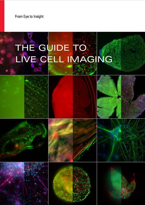
Download The Guide to Live Cell Imaging

In life science research, live cell imaging is an indispensable tool to visualize cells in a state as in vivo as possible.

LEARN MORE

Live cell imaging related articles

Read our latest articles about Live Cell Imaging

The knowledge portal of Leica Microsystems offers scientific research and teaching material on the subjects of microscopy. The content is designed to support beginners, experienced practitioners and scientists alike in their everyday work and experiments.

More Articles
An 8-color spectral unmixing result from a hyperspectral SRS (stimulated Raman scattering) dataset, showing the biochemically distinct structures of a fresh, untreated apple slice.

How to Prepare Samples for Stimulated Raman Scattering (SRS) imaging

Find here guidelines for how to prepare samples for stimulated Raman scattering (SRS), acquire images, analyze data, and develop suitable workflows. SRS spectroscopic imaging is also known as SRS…
Spheroid stained with Cyan: Dapi nuclear countertain; Green AF488 Involucrin; Orange AF55 Phalloidin Actin; Magenta AF647 CK14.

Notable AI-based Solutions for Phenotypic Drug Screening

Learn about notable optical microscope solutions for phenotypic drug screening using 3D-cell culture, both planning and execution, from this free, on-demand webinar.

Coherent Raman Scattering Microscopy Publication List

CRS (Coherent Raman Scattering) microscopy is an umbrella term for label-free methods that image biological structures by exploiting the characteristic, intrinsic vibrational contrast of their…
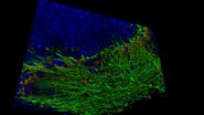
Mouse cortical neurons. Transgenic GFP (green). Image courtesy of Prof. Hui Guo, School of Life Sciences, Central South University, China

How Microscopy Helps the Study of Mechanoceptive and Synaptic Pathways

In this podcast, Dr Langenhan explains how microscopy helps his team to study mechanoceptive and synaptic pathways, their challenges, and how they overcome them.
Patch pipette touching a murine hippocampal neuron. Image courtesy of A. Aguado, Ruhr University Bochum, Germany.

What is the Patch-Clamp Technique?

This article gives an introduction to the patch-clamp technique and how it is used to study the physiology of ion channels for neuroscience and other life-science fields.
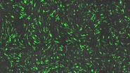
Phase-contrast image of a MDCK-cell culture and its respective confluency measured by the Mateo TL microscope.

How to Determine Cell Confluency with a Digital Microscope

This article shows how to measure cell confluency in an easy and consistent way with Mateo TL, increasing confidence in downstream experiments.
Microscope image of cultured cells at the bottom of a dish.

How to do a Proper Cell Culture Quick Check

In order to successfully work with mammalian cell lines, they must be grown under controlled conditions and require their own specific growth medium. In addition, to guarantee consistency their growth…
Combining spectrally resolved detection and fluorescence lifetime multiplexing

Live-Cell Fluorescence Lifetime Multiplexing Using Organic Fluorophores

On-demand video: Imaging more subcellular targets by using fluorescence lifetime multiplexing combined with spectrally resolved detection.

Harnessing Microfluidics to Maintain Cell Health During Live-Cell Imaging

VIDEO ON DEMAND - In this episode of MicaCam, we will use microfluidics to explore the effect of shear stress on cell morphology, examine the effect of nutrient replenishment on cellular growth during…
Living HeLa cells stained with WGA-488 (yellow), SPY-Actin (cyan), and SiR-Tubulin (magenta). Instant Computational Clearing (ICC) was applied.

How to Perform Dynamic Multicolor Time-Lapse Imaging

Live-cell imaging sheds light on diverse cellular events. As many of these events have fast dynamics, the microscope imaging system must be fast enough to record every detail. One major advantage of…
Background image
Scroll to top