Microscopy in Pathology
Microscopy in Pathology
Comparison table of DM1000 - DM3000 microscopes
| DM1000 LED | DM2000 & DM2000 LED | DM2500 & DM2500 LED | DM3000 & DM3000 LED | |
| Occasional use | x | - | - | - |
| Medium to high workload | - | x | x | x |
| Microscope height and height & viewing angle adjustable with eyepieces | x | x | x | x |
| Control knobs in proximity with symmetrical layout | - | x | x | x |
| Adjustable stage height and focus controls | x | x | x | x |
| Adjustable torque for focus knobs | - | x | x | x |
| Multiple viewing system for simultaneous observation by several users | - | - | x | - |
| Quick objective change in switching mode | - | - | - | x |
| Automated lighting management | - | - | - | x |
| Compact and robust design | x | x | x | x |
| Go to product page | Go to product page | Go to product page | Go to product page |
x = included, - = not available
Challenges related to microscopy in pathology
Pathologists need to prepare specimen slides for examination and ensure the accuracy and reliability of diagnoses, typically with strict deadlines and time constraints. They must ensure an efficient, fast processing of specimens to meet the demands of doctors treating patients in hospitals and clinics.
At times pathologists spend hours examining many specimens with a microscope. This demanding work style can lead to discomfort and repetitive strain injury (RSI) due to an uncomfortable posture while performing recurrent motions, such as adjusting the stage and focus position as well as changing objectives. Additionally, looking through the eyepieces over long periods of time with large fluctuations in light intensity can cause eyestrain.
Pathologists are in need of a solution which enables them to do microscopic examination with increased comfort and efficiency.
Advantages of the Pathology Solution Suite
Work in comfort, reduce the risk of strain,
and achieve an improved efficiency during microscopic examination.
Maintain a comfortable posture with the shoulders and spine properly aligned and attain an optimal hand positioning due to the symmetrical layout of the adjustable ergonomic stage and focus controls. The knobs are positioned on the microscope at an equal distance from the user.
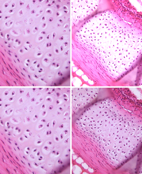
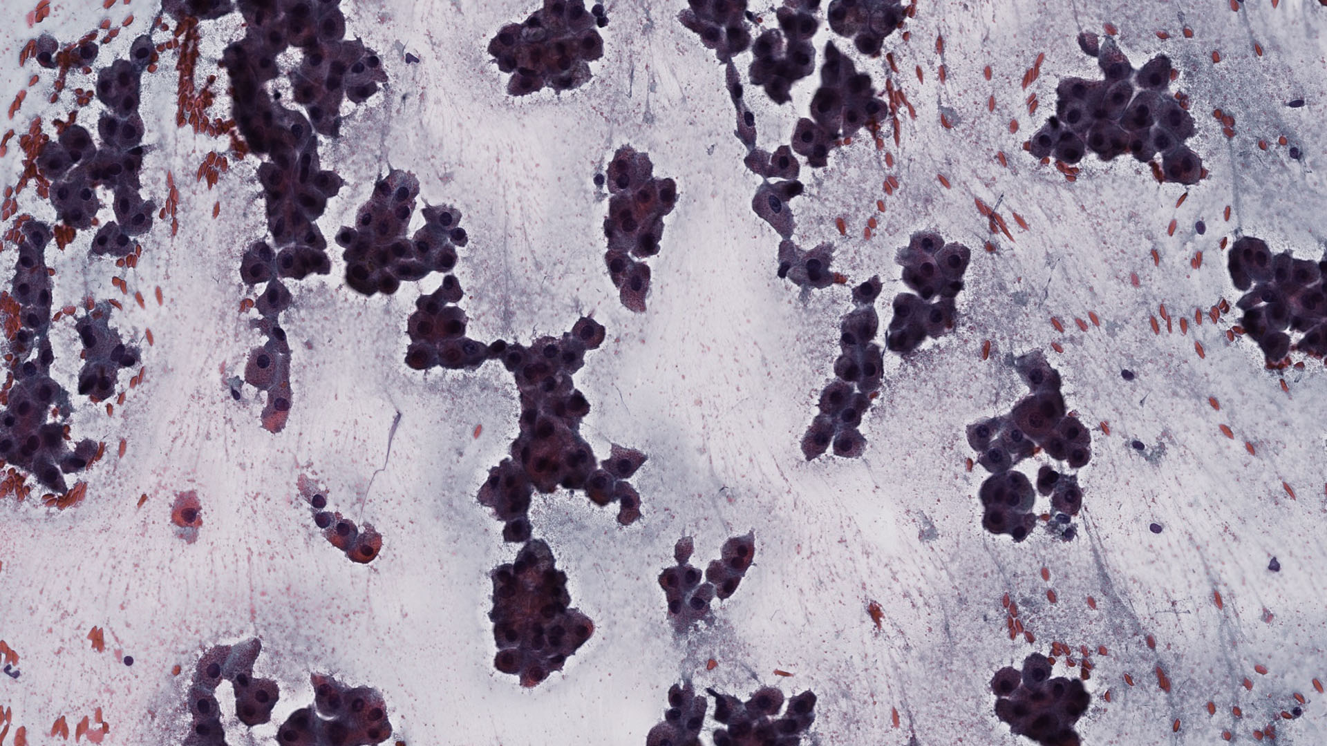
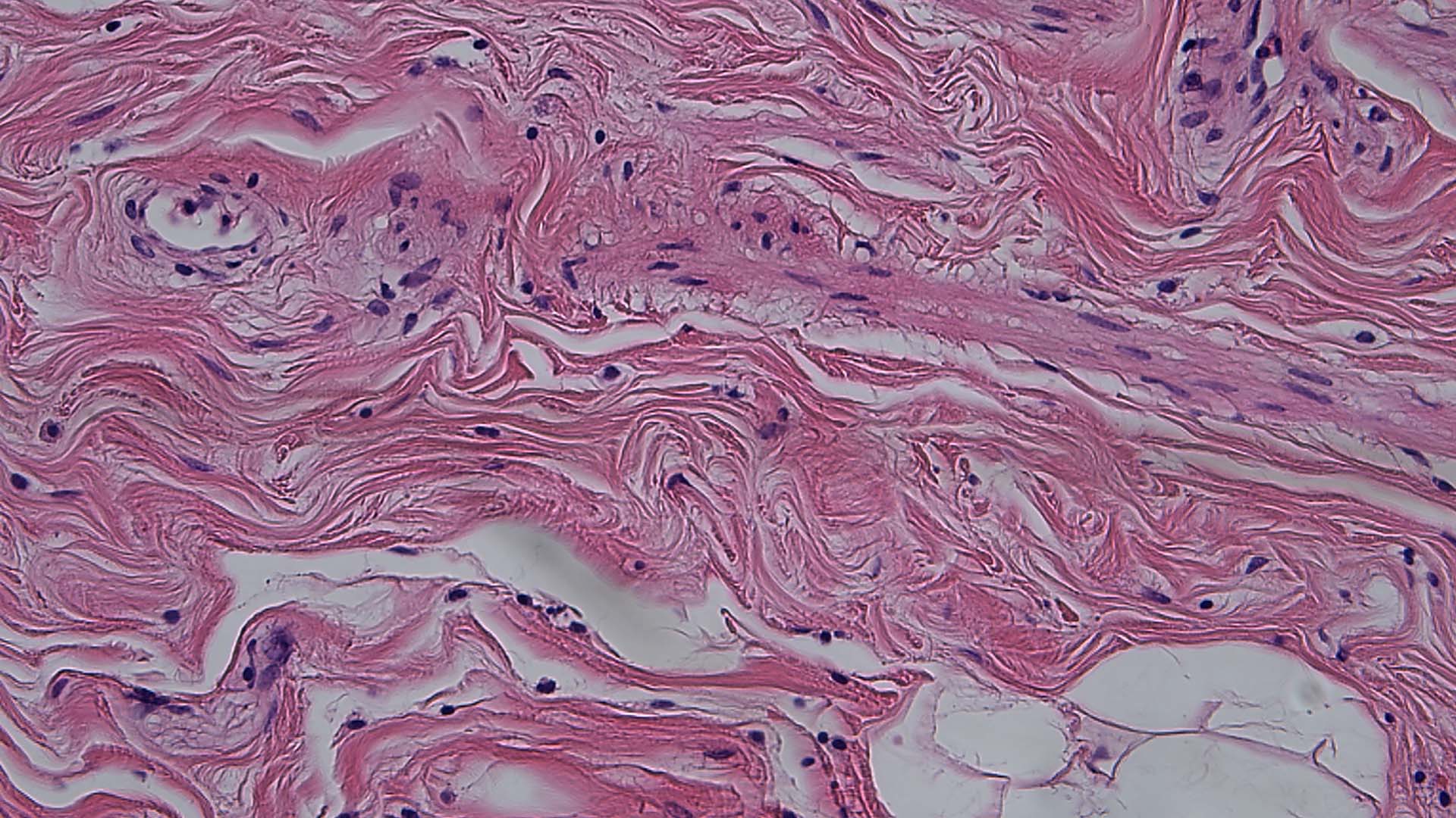
Reduce the risk of eyestrain thanks to a balanced light intensity. The brightness does not need to be continuously adjusted in order to prevent large fluctuations in light intensity.
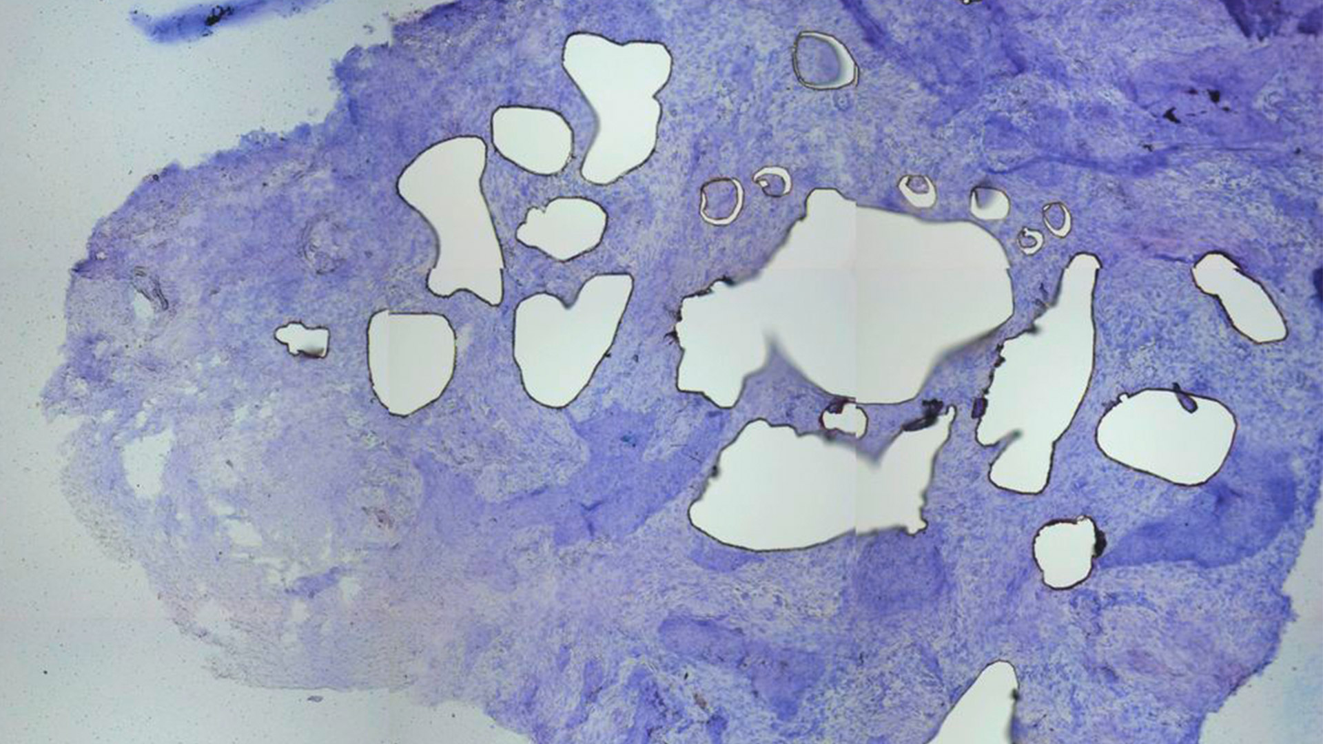
Picture is a simulation of light balancing with an image showing a trachea sample. Result after objective change from high (left) to low (right) magnification: Top right without light balancing, bottom right with light balancing.
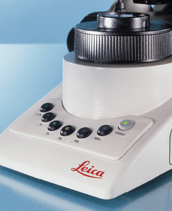
More efficient workflow thanks to minimal repetitive motions and fast objective change with the toggle mode. The knobs for the focus and stage control are in close proximity.
Customer experiences with a DM3000 microscope for clinical applications
Trudi de Jong and Marianne Noordanus, both from Rotterdam, describe how a DM3000 microscope helps them to perform their clinical microscope work more comfortably and efficiently.
Courtesy of:
Trudi de Jong, Erasmus MC academic hospital Rotterdam, the Netherlands, Hematology
Marianne Noordanus, Star-MDC, Medisch Diagnostisch Centrum, Rotterdam (The Netherlands) Microbiology
Frequently Asked Questions Pathology
Cameras are used for documentation, displaying of images on a larger screen, and showing live images for discussion among colleagues on tumor boards. They can also help pathologists with reporting of results, especially if the camera comes with software that allows annotating images and archiving them in the laboratory or hospital information system.
What pathologists need to see is important for the microscopic examination. If they need to see structures and shades of color in stained specimens, they will look at specimens in brightfield. If they need to identify the structures of cells and tissues in unstained specimens, they will use phase contrast. For more information, please refer to the Science Lab article Factors to Consider when Selecting Clinical Microscopes.
There are color cameras and monochrome cameras. Color cameras are ideally suited for pathology applications as they can visualize minute differences in staining and give pathologists a wealth of information about the specimen. Monochrome cameras are ideally suited for fluorescence applications such as FISH (fluorescence in situ hybridization). For more information, you may find this Science Lab article interesting: Clinical Microscopy: Considerations on Camera Selection.
