STELLARIS Cryo
Konfokalmikroskope
Produkte
Startseite
Leica Microsystems
STELLARIS Cryo Konfokal-Licht-Mikroskop
Lesen Sie unsere neuesten Artikel

New Imaging Tools for Cryo-Light Microscopy
New cryo-light microscopy techniques like LIGHTNING and TauSense fluorescence lifetime-based tools reveal structures for cryo-electron microscopy.

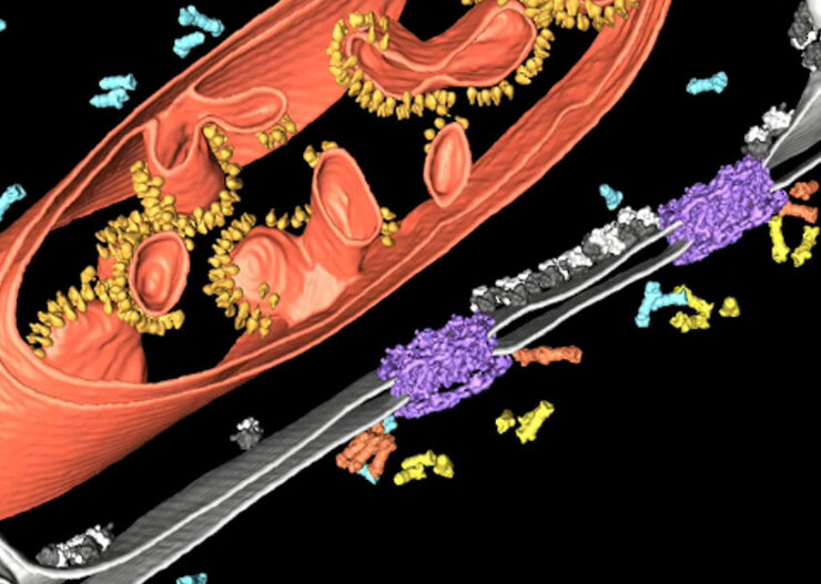
How to Target Fluorescent Structures in 3D for Cryo-FIB Milling
This article describes the major steps of the cryo-electron tomography workflow including super-resolution cryo-confocal microscopy. We describe how subcellular structures can be precisely located in…

Precise 3D Targeting for EM Imaging - Access What Matters
Find out how the seamless cryo-electron tomography workflow Coral Cryo uses confocal super resolution to target your structure of interest more precisely.

The Cryo-CLEM Journey
This article describes the Cryo-CLEM technology and the benefits it can provide for scientists. Additionally, some scientific publications are highlighted.
Recent developments in cryo electron…

Targeting Active Recycling Nuclear Pore Complexes using Cryo Confocal Microscopy
In this article, how cryo light microscopy and, in particular cryo confocal microscopy, is used to improve the reliability of cryo EM workflows is described. The quality of the EM grids and samples is…

Advancing Cell Biology with Cryo-Correlative Microscopy
Correlative light and electron microscopy (CLEM) advances biological discoveries by merging different microscopes and imaging modalities to study systems in 4D. Combining fluorescence microscopy with…

Crystal Clear Cryo Light-microscopy Images
This article describes how computational clearing of cryo light microscopy images improves the identification of cellular targets for cryo electron-microscopy.
Improve Cryo Electron Tomography Workflow
Leica Microsystems and Thermo Fisher Scientific have collaborated to create a fully integrated cryo-tomography workflow that responds to these research needs: Reveal cellular mechanisms at…
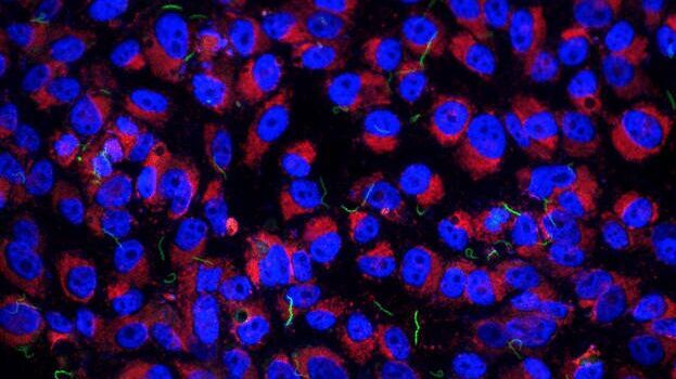
Imaging of Host Cell-bacteria Interactions using Correlative Microscopy under Cryo-conditions
Pathogenic bacteria have developed intriguing strategies to establish and promote infections in their respective hosts. Most bacterial pathogens initiate infectious diseases by adhering to host cells…
Anwendungsbereiche
Kryo-Elektronen-Tomographie
Cryo-electron tomography (CryoET) is used to resolve biomolecules within their cellular environment down to an unprecedented resolution below one nanometer.
Fortgeschrittene Mikroskopietechniken
Moderne Mikroskopietechniken umfassen sowohl hochauflösende als auch superauflösende Bildgebungsverfahren. Diese Techniken werden in erster Linie eingesetzt, um biologische Vorgänge mit extrem hoher…
Korrelative Licht- und Elektronenmikroskopie (CLEM)
Coral Workflows von Leica Microsystems unterstützt Nutzer dabei, Daten aus der Fluoreszenz- und Elektronenmikroskopie zu korrelieren (CLEM).
