Corporate Communications

Leica Microsystems entwickelt und fertigt Mikroskope und wissenschaftliche Instrumente für die Analyse von Mikro- und Nanostrukturen.
Wir bieten wissenschaftliches Forschungs- und Lehrmaterial zu den Themen der Mikroskopie an. Die Inhalte sind so gestaltet, dass sie Anfänger, erfahrene Praktiker und Wissenschaftler gleichermaßen bei ihrer täglichen Arbeit und ihren Experimenten unterstützen. Erkunden Sie interaktive Tutorials und Anwendungshinweise, entdecken Sie die Grundlagen der Mikroskopie ebenso wie High-End-Technologien.
Folgen Sie uns!

4 Key Benefits of 3D Digital Microscopy in Ophthalmic Surgery
3D digital visualization is rapidly transforming ophthalmic surgery. Modern 3D surgical microscopes enable surgeons to perform procedures using high-resolution digital displays rather than traditional…
, insulin SGs (orange), microtubules (red), nucleus (yellow), and plasma membrane (transparent).")
High-Pressure Freezing Protocols for Ultrastructural 3D EM
High pressure freezing (HPF) can help preserve hydrated cells and tissues close to their biological state at the moment of immobilization, supporting more reliable ultrastructural interpretation than…
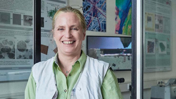
Ultramicrotome UC Enuity in Practice: Stable 15 nm Sections at ZFE
After using the UCT and UC6 ultramicrotomes, Claudia Mayrhofer calls UC Enuity a leap in stability—so robust that vibrations and temperature shifts don’t spoil sections, even with multiple users. Auto…

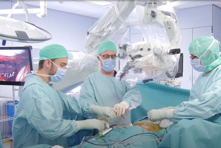
Expert Techniques for Superior Visualization in Cataract Surgery
Join renowned ophthalmic surgeons, Dr. Hussein Almuhtaseb and Mr. Simon Madge, as they share their clinical expertise and real-world surgical strategies during the 2025 Online Cataract Surgery…

Eliminating Electrostatic Interference in Laser Microdissection
Electrostatic charge in laser microdissection (LMD) causes two critical failures: samples stick to charged surfaces and are lost, or samples fly into adjacent wells and cause cross-contamination. We…

History, Developments and Trends of Microscopy in Cancer Research
Cancer is a global disease, with 18 million new cases diagnosed and 10 million cancer-related deaths worldwide in 2020. This burden is set to increase, with a projected increase in cases of ~55% by…

Researchers Insights: Microscopy in Cancer Research
Discover how imaging techniques are driving cancer research forward. In this issue, we present comprehensive multimodal studies using microscopy, as well as new directions in intraoperative cancer…

Predictive Service Prevents Downtime in Ghent
At the VIB BioImaging Core in Ghent, Belgium, researchers depend on Leica’s Stellaris 8 confocal microscope to explore the frontiers of biomedical science. When Leica’s RemoteCare system detected a…
, Tropomyosin (cardiomyocytes, red) and GFP (primordial cardiac layer, green).")
A Guide to Fluorescence Microscopy
Fluorescence microscopy uses the ability of fluorophores, dyes, or fluorescent proteins to emit light of a specific wavelength after being excited with light of a shorter wavelength. Biomolecules can…

Advances in Oncological Reconstructive Surgery
Decision making and patient care in oncological reconstructive surgery have considerably evolved in recent years. New surgical assistance technologies are helping surgeons push the boundaries of what…

Ultramicrotomy eBook: Targeting, Trimming & Alignment
Ultramicrotomy is evolving rapidly, and today’s microscopes demand high‑quality sections, precise targeting, and reproducible workflows. This eBook brings together expert application notes, automated…
 case.")
Flexibility and Efficiency in Minimally Invasive Spine Surgery
According to Prof. Alex Alfieri, Chief Physician and Head of clinic for Neurosurgery and Spinal surgery at the Cantonal Hospital Winterthur, Minimally invasive spine surgery (MISS) is transforming…
 labeled with membrane-permeable calcein, high-pressure frozen in salt water using EM ICE.")
High-Pressure Freezing for Organoids: Cryo CLEM & FIB Lift Out
Master cryo EM workflow steps for challenging 3D samples: when to choose HPF vs. plunge freezing, reproducible blotting/ice control, contamination aware transfers, Cryo CLEM 3D targeting in organoids,…
 and astrocytes (green) in a cortical spheroid derived from human induced pluripotent stem cells.")
Guide to Live-Cell Imaging
For a wide range of applications in various research fields of life science, live-cell imaging is an indispensable tool for visualizing cells in a state as close to in vivo, i.e. living and active, as…

A Larger 3D Area in Focus for Neurosurgical and Ophthalmic Microscopes
Neurosurgeons and ophthalmologists deal with delicate structures, deep or narrow cavities and tiny structures with vitally important functions. Seeing a clear, large 3D area of the surgical field in…

Factors to Consider When Selecting a Research Microscope
An optical microscope is often one of the central devices in a life-science research lab. It can be used for various applications which shed light on many scientific questions. Thereby the…

Advanced Visualization in Head and Neck Reconstructive Surgery
PRS surgeons and maxillofacial specialists face unique challenges in reconstructive procedures, particularly when operating in deep, narrow cavities and working with delicate tissues. Achieving…
Advanced Visualization: Transforming Minimally Invasive Spine Surgery
Over the past decade, spine surgery has evolved rapidly with the adoption of minimally invasive surgery (MISS) techniques. These approaches reduce surgical trauma, speed up recovery, and help improve…

Advanced Visualization in ENT Surgery: Prof. Darrouzet’s Expert Insights
Otologic surgery demands exceptional precision when working within the confined spaces of the middle and inner ear. Procedures such as tympanoplasty, mastoidectomy, and cochlear implant placement…

AI meets Deep Visual Proteomics (DVP) to Advance Disease Research
In this webinar, Dr. Andreas Mund will introduce a cutting-edge platform that merges Deep Visual Proteomics (DVP) with AI-powered pathology models, enabling high-resolution mapping of key regions in…
 and co-stained for nuclear DNA (Hoechst 33342), microtubules (Alexa 555) and F-actin (ATTO 643). Image was captured on Mateo FL.")
Microscopy and AI Solutions for 2D Cell Culture
This eBook explores the integration of microscopy and AI technologies in 2D cell culture workflows. It highlights how traditional imaging methods—such as brightfield, phase contrast, and…
.")
Focus on Long-Term Imaging in 3D with Light Sheet Microscopy
Long-term 3D imaging reveals how complex multicellular systems grow and develop and how cells move and interact over time, unlocking critical insights into development, disease, and regeneration.…

Polarizing Microscope Image Gallery
How polarization microscope images can be used for analysis is shown in this gallery. Polarized light microscopy (also known as polarizing microscopy) is an important method for different fields and…
 and tubulin (magenta), acquired using Viventis Deep. Courtesy of Akanksha Jain, Treutlein Lab ETH-DBSSE Basel (Switzerland).")
Faster & Deeper Insights into Organoid and Spheroid Models
Gain deeper, more translatable, insights into organoid and spheroid models for drug discovery and disease research by overcoming key imaging challenges. In this eBook, explore advanced microscopy…

A Guide to C. elegans Research – Working with Nematodes
Efficient microscopy techniques for C. elegans research are outlined in this guide. As a widely used model organism with about 70% gene homology to humans, the nematode Caenorhabditis elegans (also…
 at Leica Microsystems.")
How a Breakthrough in Spatial Proteomics Saved Lives
Toxic epidermal necrolysis (TEN) is a rare but devastating reaction to common medications like antibiotics or gout treatments. It begins innocuously, often as a rash, but can escalate rapidly into…
 performing ear, nose and throat (ENT) surgery using the MyVeo surgical visualization headset.")
A Microvascular Surgeon’s View: How MyVeo Transforms Visualization
In this article, Dr. Andrew T. Huang, MD, FACS, otolaryngologist and a head and neck reconstructive surgeon, shares how digital 3D surgical visualization with the MyVeo headset from Leica Microsystems…

Development and Derisking of CRISPR Therapies for Rare Diseases
This on-demand presentation by Dr. Fyodor Urnov and Dr. Sadik Kassim, originally delivered at ASGCT 2025, focused on a critical challenge in genetic medicine: how to scale CRISPR therapies from…

Integrated Serial Sectioning and Cryo-EM Workflows for 3D Biological Imaging
This on-demand webinar explores how integrated tools can support electron microscopy workflows from sample preparation to image analysis. Experts Andreia Pinto, Adrian Boey, and Hoyin Lai present the…

Revealing Sodium Battery Degradation via Cryo-EM and CryoFIB
Explore how cryogenic electron microscopy and focused ion beam techniques uncover the intrinsic structure of sodium battery interfaces. This webinar presents a new degradation model based on separator…
