Corporate Communications

Leica Microsystems entwickelt und fertigt Mikroskope und wissenschaftliche Instrumente für die Analyse von Mikro- und Nanostrukturen.
Wir bieten wissenschaftliches Forschungs- und Lehrmaterial zu den Themen der Mikroskopie an. Die Inhalte sind so gestaltet, dass sie Anfänger, erfahrene Praktiker und Wissenschaftler gleichermaßen bei ihrer täglichen Arbeit und ihren Experimenten unterstützen. Erkunden Sie interaktive Tutorials und Anwendungshinweise, entdecken Sie die Grundlagen der Mikroskopie ebenso wie High-End-Technologien.
Folgen Sie uns!

Image Gallery: THUNDER Imager
To help you answer important scientific questions, THUNDER Imagers eliminate the out-of-focus blur that clouds the view of thick samples when using camera-based fluorescence microscopes. They achieve…

Plastische und rekonstruktive Chirurgie: Warum ein Mikroskop benutzen?
Verfahren der plastischen und rekonstruktiven Chirurgie können heikel sein. Visualisierungslösungen spielen eine wichtige Rolle, da sie es ermöglichen, die Operation unter bestmöglichen Bedingungen…

Minimally Invasive Spine Surgery: Improving Precision and Accuracy with Microscopes
Spine surgery is extremely delicate and requires extensive training and experience. Innovative visualization technologies can also help achieve better outcomes allowing to see more and have a clearer…

Modellorganismen in der Forschung
Modellorganismen sind Spezies, mit denen Forscher bestimmte biologische Vorgänge untersuchen. Sie haben genetische Ähnlichkeiten mit Menschen und werden häufig in Forschungsbereichen wie Genetik,…

Mit LIGHTNING das Maximum an Informationen aus Ihrer Probe erhalten
LIGHTNING ist ein adaptiver Prozess zur Extraktion von Bildinformationen, bei dem vollautomatisch anderweitig nicht sichtbare Strukturen und feine Details sichtbar gemacht werden. Im Gegensatz zu…
Advanced Techniques in Cataract and Refractive Surgery
In this webinar Dr. Thompson and Dr. Moshirfar will explain how Leica microscopes aid in procedures such as Centration of Multifocal IOLs and corneal inlays such as Kamra and Lenticular Grafts used in…

Virologie
Liegt Ihr Forschungsschwerpunkt auf Virusinfektionen und -krankheiten? Erfahren Sie, wie Sie mit Lösungen für Bildgebung und Probenvorbereitung von Leica Microsystems mehr Erkenntnisse in der…

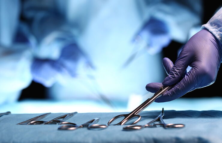
Surgical Microscopes: Key Factors for OR Nurses
Operating room (OR) nurses are vital to the surgery process. An OR Nurse Manager explains the key surgical microscope features to facilitate their work.

Computational Clearing - Enhance 3D Specimen Imaging
This webinar is designed to clarify crucial specifications that contribute to THUNDER Imagers' transformative visualization of 3D samples and improvements within a researcher's imaging-related…

STELLARIS White Light Lasers
When it comes to choosing fluorescent probes for your multi-color experiments, you shouldn’t have to compromise. Now you can advance beyond conventional excitation sources that limit your fluorophore…
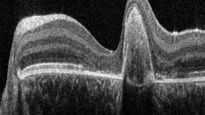
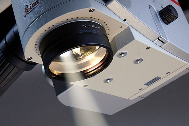
A Guide to OCT
Leica Optical Coherence Tomography (OCT) systems support ophthalmologists, ophthalmic surgeons, and researchers with easy-to-use, high-quality imaging technology.

Krebsforschung
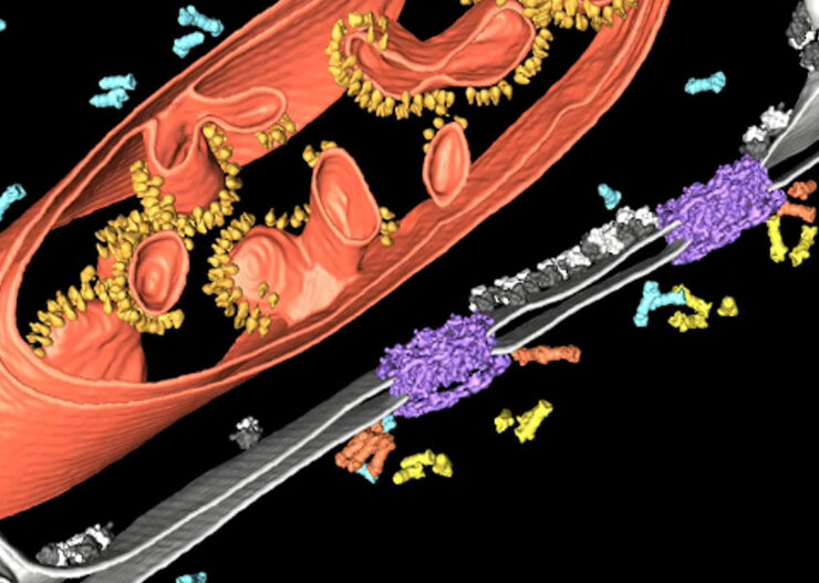
Krebs ist eine komplexe und heterogene Krankheit, die durch Zellen verursacht wird, denen die Wachstumsregulation fehlt. Genetische und epigenetische Veränderungen in einer Zelle oder einer Gruppe von…
, and trafficking vesicles labeled with CF594 (cyan - Biotium).")
A Guide to Super-Resolution
Find out more about Leica super-resolution microscopy solutions and how they can empower you to visualize in fine detail subcellular structures and dynamics.
How to Drape an Overhead Surgical Microscope
The tutorial features the Leica ARveo digital Augmented Reality microscope for complex neurosurgery. The procedure also applies to the Leica M530 OHX, OH6, OH5 and OH4.

How to Drape a Surgical Microscope
Before performing surgical procedures, it is important to drape the surgical microscope to ensure sterile working conditions. At Leica, we are committed to helping you with your surgical practice. In…
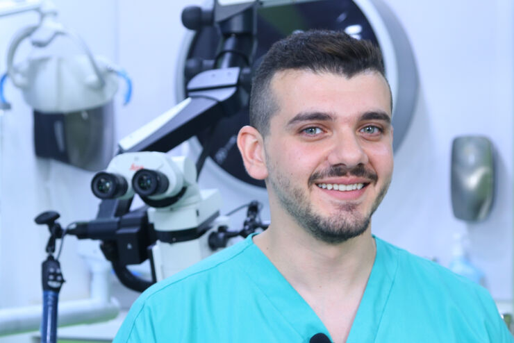
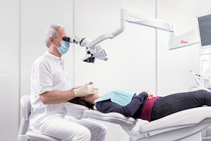
Overcoming Complexities in Microdentistry
Dr. Salam Abu Arqub, from the Smile Engineer Dental Center in Amman, Jordan, has been using Leica dental microscopes for three years for all procedures performed at the clinic. He shared his…

THUNDER Imagers: High Performance, Versatility and Ease-of-Use for your Everyday Imaging Workflows
This webinar will showcase the versatility and performance of THUNDER Imagers in many different life science applications: from counting nuclei in retina sections and RNA molecules in cancer tissue…
Improve Cryo Electron Tomography Workflow
Leica Microsystems and Thermo Fisher Scientific have collaborated to create a fully integrated cryo-tomography workflow that responds to these research needs: Reveal cellular mechanisms at…

Improve 3D Cell Biology Workflow with Light Sheet Microscopy
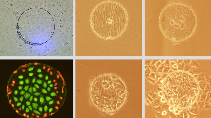
Understanding the sub-cellular mechanisms in carcinogenesis is of crucial importance for cancer treatment. Popular cellular models comprise cancer cells grown as monolayers. But this approach…
Live Cell Isolation by Laser Microdissection
Laser microdissection is a tool for the isolation of homogenous cell populations from their native niches in tissues to downstream molecular assays. Beside its routine use for fixed tissue sections,…
Erfolgreiche endodontische Behandlung mit Dental-Operationsmikroskopen
In der Endodontie hängt die zielgerichtete Behandlung nicht nur von den technischen Fähigkeiten und dem Wissen des Zahnarztes ab. Es kommt auch auf die klare, detaillierte Visualisierung des…
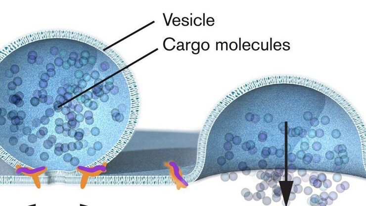
Nobel Prize 2013 in Physiology or Medicine for Discoveries of the Machinery Regulating Vesicle Traffic
On October 7th 2013, The Nobel Assembly at Karolinska Institutet has decided to award The Nobel Prize in Physiology or Medicine 2012 jointly to James E. Rothman, Randy W. Schekman and Thomas C. Südhof…
