Kontaktieren Sie uns

Rolf T. Borlinghaus , Dr.

Rolf T. Borlinghaus

Rolf Borlinghaus wurde 1956 in Grötzingen, Deutschland, geboren. Nach seinem Diplom in Biologie arbeitete er im Labor von Peter Läuger im Fachbereich Biophysik der Universität Konstanz an elektrogenen Schritten der Na/K-ATPase durch laserinduzierte Freisetzung von ATP aus einer eingeschlossenen Verbindung, von wo aus er 1988 zum Dr.rer.nat. befördert wurde. 1990 begann er als Produktmanager für Fluoreszenz- und konfokale Forschungsmikroskope bei Carl Zeiss in Oberkochen zu arbeiten. 1997 wechselte er zu Leica (damals Leica Lasertechnik, Heidelberg), um sich dieser Herausforderung zu stellen. Aus persönlichen Gründen hat Rolf Borlinghaus 2007 seine Führungsaufgaben abgegeben und unterstützte als Senior Scientist mit einer halben Stelle die konfokale Marketinggruppe. Die andere Hälfte widmet er den Bereichen Beziehungen, Lebensmittel und Botanik.

Springer-Publikationen:

2017 Die Lichtblattmikroskopie (DE)

2017 The White Confocal (EN)

2016 Konfokale Mikroskopie in Weiß (DE)

2016 Unbegrenzte Lichtmikroskopie (DE)

Rolf Borlinghaus ging im Dezember 2020 in den Ruhestand.

Nachdem wir im Mai 2021 erfahren haben, dass Rolf Borlinghaus verstorben ist, möchten seine Kollegen ihren Dank und ihre Anerkennung für sein langjähriges Engagement für Leica Microsystems und seine Kunden zum Ausdruck bringen. Seine zahlreichen Beiträge zur Mikroskopie, die auf Science Lab und in wissenschaftlichen Zeitschriften und Büchern veröffentlicht wurden, werden in Erinnerung bleiben.

Going Beyond Deconvolution

Widefield fluorescence microscopy is often used to visualize structures in life science specimens and obtain useful information. With the use of fluorescent proteins or dyes, discrete specimen…

Drosophila Testis Niche Stem Cells – Three Color Computational Clearing

Differentiated living beings such as humans, but also a fruit fly or a plant, possess not only the differentiated cells which form specific tissues, but also those cells whose fate is not yet (or only…

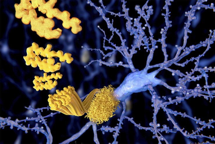
Alzheimer Plaques: fast Visualization in Thick Sections

More than 60% of all diagnosed cases of dementia are attributed to Alzheimer’s disease. Typical of this disease are histological alterations in the brain tissue. So far, there is no cure for this…
Zebrafish Whole Brain imaging with Leica SP8 spectral confocal laser scanning microscope

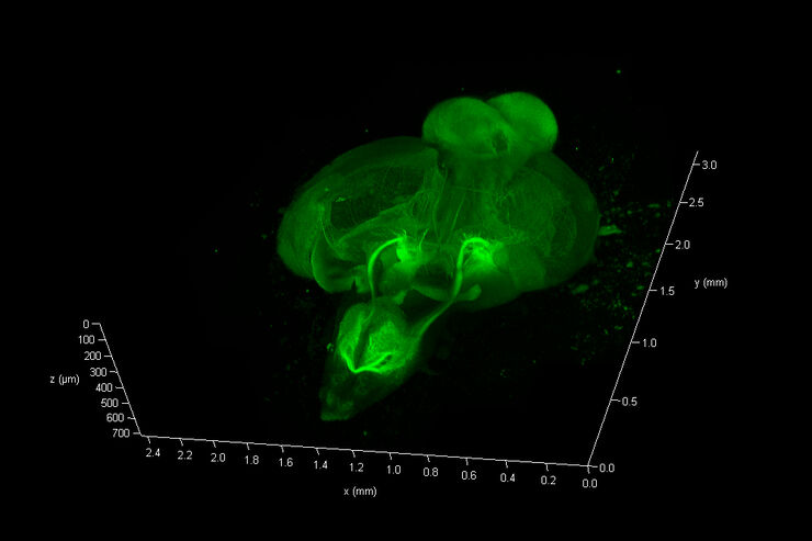
Zebrafish Brain - Whole Organ Imaging at High Resolution

Structural information is key when one seeks to understand complex biological systems, and one of the most complex biological structures is the vertebrate central nervous system. To image a complete…

What is a Spectral Detector (SP Detector)?

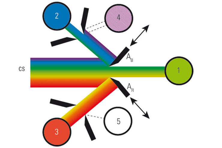
The SP detector from Leica Microsystems denotes a compound detection unit for point scanning microscopes, in particular confocal microscopes. The SP detector splits light into up to 5 spectral bands.…

What is a Resonant Scanner?

A resonant scanner is a type of galvanometric mirror scanner that allows fast image acquisition with single-point scanning microscopes (true confocal and multiphoton laser scanning). High acquisition…

What is a Field-of-View Scanner?

A field-of-view scanner is an assembly of galvanometric scanning mirrors used in single-point confocal microscopes that offer the correct optical recording of large field sizes. The field-of-view…

Resolved Field Number (RFN)

The field number (FN) for optical microscopes indicates the field of view (FOV). It corresponds to the area in the intermediate image that is observable through the eyepieces. Although, we cannot…

What is a Tandem Scanner?

A Tandem Scanner is an assembly of two different types of scanning together in one system for true confocal point scanning. The Tandem Scanner consists of a three-mirror scanning base with the…

Which Sensor is the Best for Confocal Imaging?

The Hybrid Photodetectors (HyD) are! Why that is the case is explained in this short Science Lab article.
Multiphoton microscopy of an unstained mouse skin section acquired using the 4Tune detector.

Mission Impossible Accomplished: Tunable Colors for Non-descanning Detection

Leica Microsystems’ 4Tune detector, the key component of the SP8 DIVE Deep In Vivo Explorer, provides spectrally tunable image recording with non-descanning detection. An innovative solution for…
„Confetti-Mouse“, taken with the 4Tune spectral detector of Leica’s Deep in-vivo Explorer SP8 DIVE.

Laser Beam Shaping for Multicolor Multiphoton Microscopy

Multiphoton Microscopy is one of the current hot topics in life science research. The new Leica TCS SP8 DIVE from Leica Microsystems presents a series of beneficial new innovations, including a freely…

Primary Beam Splitting Devices for Confocal Microscopes

Current fluorescence microscopy employs incident illumination which requires separation of illumination and emission light. The classical device performing this separation is a color-dependent beam…
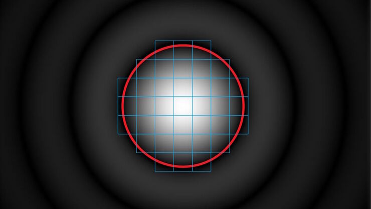
Airy-Muster in der Fokusebene

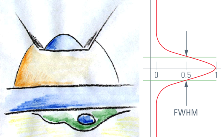
Was macht das Pinhole im konfokalen Mikroskop?

Wer ein konfokales Mikroskop benutzt und über die Eigenschaften und Parameter eines solchen Gerätes diskutiert, wird notwendigerweise über das Pinhole reden – und wie dessen Größe sich auf das…

Vom Licht zur Erleuchtung: Sensoren und Messverfahren in der konfokalen Mikroskopie

In diesem Beitrag werden die wichtigsten Sensoren kurz vorgestellt, die in der konfokalen Mikroskopie verwendet werden. Mit konfokaler Mikroskopie ist hier „True Confo-cal Scanning“ gemeint, also das…
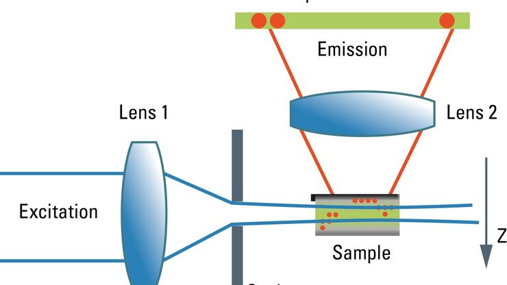
Schematic graph of the light path in a Spalt-Ultramikroskop.

Confocal and Light Sheet Imaging with STELLARIS DLS

Optical imaging instrumentation can magnify tiny objects, zoom in on distant stars and reveal details that are invisible to the naked eye. But it notoriously suffers from an annoying problem: the…
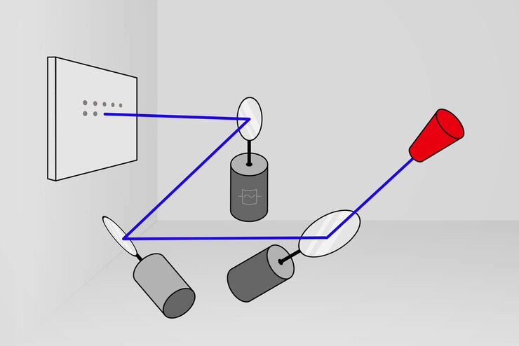
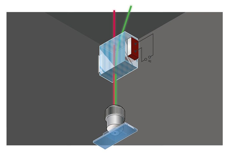
Acousto-optics, sketch

Acousto Optics in True Confocal Spectral Microscope Systems

Acousto-optical elements have successfully replaced planar filters in many positions. The white confocal, regarded as the fully spectrally tunable confocal microscope, was not possible without this…

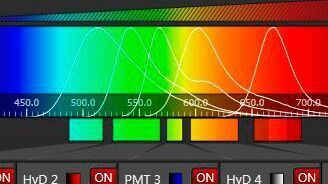
Spectral Detection – How to Define the Spectral Bands that Collect Probe-specific Emission

To specifically collect emission from multiple probes, the light is first separated spatially and then passes through a device that defines a spectral band. Classically, this is a common glass-based…
Scroll to top