Leica Microsystems

Leica Microsystems

Leica Microsystems ist ein weltweit führender Hersteller von Mikroskopen und wissenschaftlichen Instrumenten. Im 19. Jahrhundert als Familienunternehmen gegründet, war die Geschichte des Unternehmens auf dem Weg zum Weltkonzern von beispielloser Innovation geprägt.

Die traditionell enge Zusammenarbeit mit der Wissenschaft ist der Schlüssel zur Innovationstradition von Leica Microsystems, die auf die Ideen der Anwender zurückgreift und auf deren Bedürfnisse zugeschnittene Lösungen schafft. Auf globaler Ebene ist Leica Microsystems in drei Divisionen gegliedert, die alle zu den führenden Anbietern in ihrem jeweiligen Bereich gehören: Life Science, Industrie und Medizin.

Das Unternehmen ist in über 100 Ländern mit 6 Produktionsstandorten in 5 Ländern, Vertriebs- und Serviceorganisationen in 20 Ländern und einem internationalen Händlernetz vertreten. Der Hauptsitz des Unternehmens liegt in Wetzlar, Deutschland.

https://www.leica-microsystems.com/de

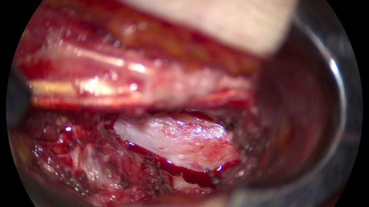
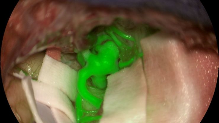
Aneurysm shown with GLOW800 AR fluorescence application. Image courtesy of Prof. Jacques Guyotat, Hôpital Neurologique Pierre Wertheimer, Lyon

How AR Fluorescence Imaging Supports Neurovascular Surgery

In this article, we explain how fluorescence imaging works in vascular neurosurgery and explain the benefits of the GLOW800 Augmented Reality fluorescence application.
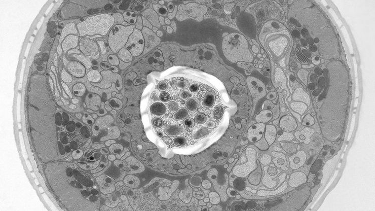
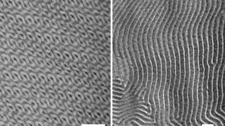
TEM micrographs of polymer sections. Left: Poly(styrene)-b-poly(isoprene). Right: Poly(styrene)-b-poly(methyl methacrylate).

Ultramicrotome Sectioning of Polymers for TEM Analysis

We demonstrate the capabilities of the UC Enuity ultramicrotome from Leica Microsystems for preparing ultrathin sections of polymer samples under both ambient and cryogenic conditions. By presenting…
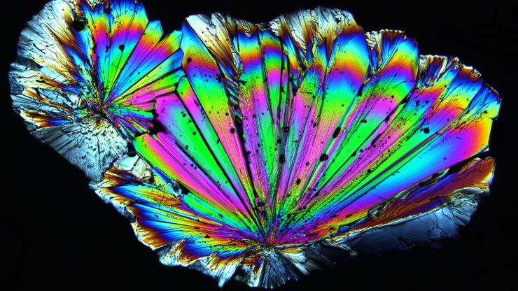
Radially grown sugar crystals which have been imaged with a Leica microscope using circular polarized light.

Polarizing Microscope Image Gallery

How polarization microscope images can be used for analysis is shown in this gallery. Polarized light microscopy (also known as polarizing microscopy) is an important method for different fields and…
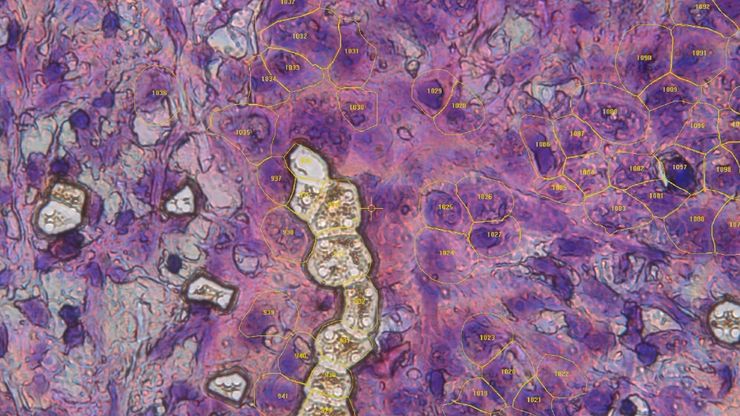
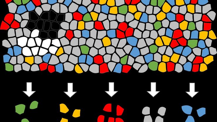
Artificial Intelligence (AI) segmentation used in conjunction with LMD to increase discovery throughput.

Biomarker Discovery with Laser Microdissection

Explore the potential of spatial proteomics workflows, such as Deep Visual Proteomics (DVP), to decipher pathology mechanisms and uncover druggable targets. Altered protein expression, abundance, or…
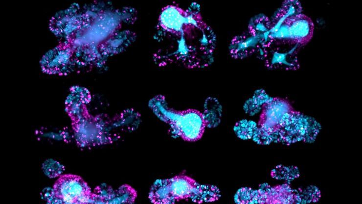
Brain organoid labeled with lamin (green) and tubulin (magenta), acquired using Viventis Deep. Courtesy of Akanksha Jain, Treutlein Lab ETH-DBSSE Basel (Switzerland).

Faster & Deeper Insights into Organoid and Spheroid Models

Gain deeper, more translatable, insights into organoid and spheroid models for drug discovery and disease research by overcoming key imaging challenges. In this eBook, explore advanced microscopy…
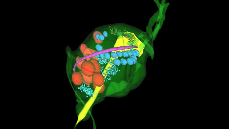
Final Segmentation of organelles in Trichomonas species. Magenta – costa, light blue – hydrogenosomes, turquoise – ER, red – vacuoles, yellow – axostyle, green – Golgi apparatus.  Sample courtesy of Isabelle Guerin-Bonne, Low Kay En, Electron Microscopy Unit, Yong Loo Lin School of Medicine, National University of Singapore. Scale bar: 1 µm.

Volume EM and AI Image Analysis

The article outlines a detailed workflow for studying biological tissues in three dimensions using volume-scanning electron microscopy (volume-SEM) combined with AI-assisted image analysis. The focus…
Image of roundworm C. elegans acquired with a M205 FA fluorescence automated stereo microscope in combination with Rottermann contrast. Areas labelled with mCherry are seen as reddish purple.

A Guide to C. elegans Research – Working with Nematodes

Efficient microscopy techniques for C. elegans research are outlined in this guide. As a widely used model organism with about 70% gene homology to humans, the nematode Caenorhabditis elegans (also…

How a Breakthrough in Spatial Proteomics Saved Lives

Toxic epidermal necrolysis (TEN) is a rare but devastating reaction to common medications like antibiotics or gout treatments. It begins innocuously, often as a rash, but can escalate rapidly into…
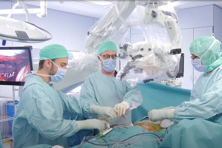
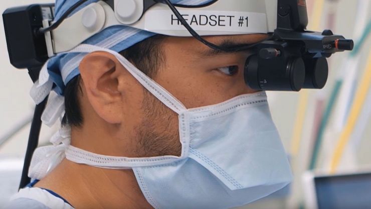
Dr. Andrew Huang, Baylor College of Medicine, in the operating room (OR) performing ear, nose and throat (ENT) surgery using the MyVeo surgical visualization headset.

A Microvascular Surgeon’s View: How MyVeo Transforms Visualization

In this article, Dr. Andrew T. Huang, MD, FACS, otolaryngologist and a head and neck reconstructive surgeon, shares how digital 3D surgical visualization with the MyVeo headset from Leica Microsystems…
Fluorescence microscopy of sectioned tissue, showing the interface between the extensor digitorum longus muscle and the common peroneal nerve in the adult rat. Regenerative peripheral nerve interface (RPNI) at 2 weeks. Image acquired using Mica. Stained for nuclei (blue), neurofilaments (green) and S100B (red). Image courtesy of Dr. Aaron Lee, Department of Bioengineering (Lab of Dr. Rylie Green), Imperial College London.

How to Image Axon Regeneration in Deep Muscle Tissue

This study highlights Dr. Aaron Lee’s research on mapping nerve regeneration in muscle grafts post-amputation. Limb loss often leads to reduced quality of life, not only from tissue loss but also due…
5 hour time-lapse maximum intensity projection of a zebrafish embryo along the z-axis at 3 days post fertilization. Left: microglia cells. Right: bright field channel. Courtesy of Prof. Francesca Peri, University of Zurich, Switzerland.

Capturing Developmental Dynamics in 3D

This application note showcases how the Viventis Deep dual-view light sheet microscope was successfully used by researchers for exploring high-resolution, long-term imaging of 3D multicellular models…
Some 2D measurements, e.g., lengths and areas, made on a PCB sample with a Leica measurement microscope using the Enersight software.

How to Select the Right Measurement Microscope

With a measurement microscope, users can measure the size and dimensions of sample features in both 2D and 3D, something crucial for inspection, QC, failure analysis, and R&D. However, choosing the…
These images illustrate the need for multiple z-slices to capture all gH2Ax foci in a given cell and get an accurate count.

Development and Derisking of CRISPR Therapies for Rare Diseases

This on-demand presentation by Dr. Fyodor Urnov and Dr. Sadik Kassim, originally delivered at ASGCT 2025, focused on a critical challenge in genetic medicine: how to scale CRISPR therapies from…
Example of calibrating a microscope at a higher magnification value using a stage micrometer.

Microscope Calibration for Measurements: Why and How You Should Do It

Microscope calibration ensures accurate and consistent measurements for inspection, quality control (QC), failure analysis, and research and development (R&D). Calibration steps are described in this…

Integrated Serial Sectioning and Cryo-EM Workflows for 3D Biological Imaging

This on-demand webinar explores how integrated tools can support electron microscopy workflows from sample preparation to image analysis. Experts Andreia Pinto, Adrian Boey, and Hoyin Lai present the…
SEM image of the full Li-NMC electrode sample, showing the two porous layers and the metal film at the center of the structure.

Revealing Sodium Battery Degradation via Cryo-EM and CryoFIB

Explore how cryogenic electron microscopy and focused ion beam techniques uncover the intrinsic structure of sodium battery interfaces. This webinar presents a new degradation model based on separator…
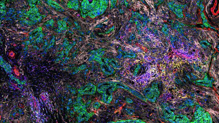
Cell DIVE multiplexed image of FFPE tissue section from human colon adenocarcinoma tissue.

Multiplexed Imaging Reveals Tumor Immune Landscape in Colon Cancer

Cancer immunotherapy benefits few due to resistance and relapse, and combinatorial therapeutic strategies that target multiple steps of the cancer-immunity cycle may improve outcomes. This study shows…
A fruit fly (Drosophila melanogaster) observed with an Ivesta 3 stereo microscope during fly pushing (sorting of the flies). The scale bar length is 1 mm. Image courtesy of M. Benton, EMBL, Heidelberg, Germany.

A Guide to Using Microscopy for Drosophila (Fruit Fly) Research

The fruit fly, typically Drosophila melanogaster, has been used as a model organism for over a century. One reason is that many disease-related genes are shared between Drosophila and humans. It is…
Cell DIVE multiplexed image of FFPE tissue section from human invasive ductal carcinoma (IDC)

AI-Powered Hi-Plex Spatial Analysis Tools for Breast Cancer Research

Breast cancer (BC) is the leading cause of cancer-related deaths in women. Investigating the tumor microenvironment (TME) is crucial to elucidate the mechanisms of tumor progression. Systematic…
Mouse brain slice which was immunostained with GFAP-A647 and imaged using a THUNDER Imager Tissue. Courtesy of H. Xu, University of Pennsylvania, Philadelphia, USA.

Neurowissenschaften

Arbeiten Sie an einem besseren Verständnis neurodegenerativer Erkrankungen oder an einer Untersuchung der Funktionen des Nervensystems? Erfahren Sie, wie Sie mit Bildgebungslösungen von Leica…
Mouse hippocampus brain slice on a grid after HPF using the “Waffle Method”.

The “Waffle Method”: High-Pressure Freeze Complex Samples

This article describes the advantages of a special high pressure freezing method, the so-called “Waffle Method”. Learn how the “Waffle Method” uses EM grids as spacers for high-pressure freezing,…
UC Enuity with Diatome diamond knife.

Mastering Polymer Sectioning with Helmut Gnaegi

When it comes to ultramicrotomy, few names carry the weight of Helmut Gnaegi. As co-founder of Diatome, a global leader in diamond knife technology, Helmut has spent decades refining the art and…

Zebrafisch-Forschung

Für optimale Ergebnisse während der Bewertung, Sortierung, Manipulation und Bildgebung von Modellorganismen ist es entscheidend feine Details und Strukturen genauestens zu erkennen. Das bildet die…
3D high-plex imaging in cancer immunology. Overview of a pancreatic tumor section in mouse model, labeled with 15 markers and imaged in one go using STELLARIS with SpectraPlex. (https://www.nature.com/articles/d42473-024-00260-7)

How to Streamline High-Plex Imaging for 3D Spatial Omics Advances

In this webinar, Dr. Julia Roberti and Dr. Luis Alvarez from Leica Microsystems introduce SpectraPlex, a new functionality integrated into the STELLARIS confocal platform for high-plex 3D spatial…
Evolved ARveo and MyVeo in Operating Room

The Guide to Augmented Reality in Microsurgery

In an era of technological advancement, Augmented Reality (AR) is rapidly transforming the medical field. In surgical microscopy, AR can display fluorescence signals as digital overlays in real-time…
Zebrafish-embryo image captured using a THUNDER Imager Tissue and live instant computational clearing.

Improving Zebrafish-Embryo Screening with Fast, High-Contrast Imaging

Discover from this article how screening of transgenic zebrafish embryos is boosted with high-speed, high-contrast imaging using the DM6 B microscope, ensuring accurate targeting for developmental…
Pancreatic Ductal Adenocarcinoma with 11 Apoptosis biomarkers shown – BAK, BAX, BCL2, BCLXL, Caspase9, CIAP1, NaKATPase, PCK26, SMAC, Vimentin, and XIAP.

Transforming Research with Spatial Proteomics Workflows

Spatial Proteomics, Nature Methods 2024 Method of the Year, is driving research advancements in cancer, immunology, and beyond. By combining positional data with high throughput imaging of proteins in…
C. elegans embedded in Lowicryl® HM20; pharynx showing red fluorescence (mCherry). The overview shows a front view onto the resin capsule formed by the bottom of a flow-through chamber of the EM AFS2. The capsule was pretrimmed manually. The blockface was trimmed automatically using the AutoTrim function of UC Enuity guided by fluorescence of the worm. Edge length of both squares in relation to the images is 250 µm.

How Fluorescence Guides Sectioning of Resin-embedded EM Samples

Electron microscopes, including transmission electron microscopes (TEM) and scanning electron microscopes (SEM), are widely utilized to gain detailed structural information about biological samples or…

Coherent Raman Scattering Microscopy Publication List

CRS (Coherent Raman Scattering) microscopy is an umbrella term for label-free methods that image biological structures by exploiting the characteristic, intrinsic vibrational contrast of their…
Ivesta 3 with integrated monitor.

Präpariermikroskope

Wenn Sie Präparationen durchführen, verbringen Sie oft viele Stunden an den Okularen eines Präpariermikroskops. Bei Leica Microsystems können Sie aus einer Vielzahl von Mikroskopen und einem…
Scroll to top