
Ciencias de la vida
Ciencias de la vida
Este es el lugar para ampliar sus conocimientos, capacidades de investigación y aplicaciones prácticas de la microscopía en diversos campos científicos. Aprenda a conseguir una visualización precisa, interpretación de imágenes y avances en la investigación. Encuentre información detallada sobre microscopía avanzada, técnicas de obtención de imágenes, preparación de muestras y análisis de imágenes. Los temas tratados incluyen la biología celular, la neurociencia y la investigación del cáncer, con especial atención a las aplicaciones e innovaciones de vanguardia.

Understanding Motor Sequence Generation Across Spatiotemporal Scales
We have developed a microscopy-based pipeline to characterize a developmentally critical behavior at the pupal stage of development, called the ecdysis sequence. We study brain-wide neuronal activity…

Benefits of TauContrast to Image Complex Samples
In this interview, Dr. Timo Zimmermann talks about his experience with the application of TauSense tools and their potential for the investigation of demanding samples such as thick samples or…

Fast, High-quality Vitrification with the EM ICE High Pressure Freezer
The EM ICE High Pressure Freezer was developed with a unique freezing principle and uses only a single pressurization and cooling liquid: liquified nitrogen (LN2). This design enables three major…

Targeting Active Recycling Nuclear Pore Complexes using Cryo Confocal Microscopy
In this article, how cryo light microscopy and, in particular cryo confocal microscopy, is used to improve the reliability of cryo EM workflows is described. The quality of the EM grids and samples is…

Investigating Synapses in Brain Slices with Enhanced Functional Electron Microscopy
A fundamental question of neuroscience is: what is the relationship between structural and functional properties of synapses? Over the last few decades, electrophysiology has shed light on synaptic…

The Power of Pairing Adaptive Deconvolution with Computational Clearing
Learn how deconvolution allows you to overcome losses in image resolution and contrast in widefield fluorescence microscopy due to the wave nature of light and the diffraction of light by optical…
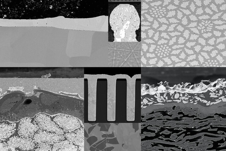
Ion Beam Milling Guide: Enhancing Surface Quality for High-Resolution Imaging and Analysis
In this article you can learn how to optimize the preparation quality of your samples by using the ion beam etching method with the EM TIC 3X ion beam milling machine. A short introduction of the…

Soluciones de recubrimiento con pulverización catódica y crío-fractura
Leica Microsystems cubre toda la gama de necesidades de recubrimiento, desde el recubrimiento a baja temperatura ambiente hasta el crío-recubrimiento de alto vacío.

Studying Pulmonary Fibrosis
The results shown in this article demonstrate that fibrotic and non-fibrotic regions of collagen present in mouse lung tissue can be distinguished better with polarized light compared to brightfield.…
