
Scienze della vita
Scienze della vita
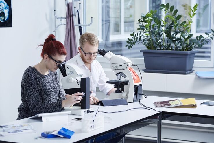
Questo è il posto giusto per ampliare le vostre conoscenze, le capacità di ricerca e le applicazioni pratiche della microscopia in vari campi scientifici. Imparate come ottenere una visualizzazione precisa, l'interpretazione delle immagini e i progressi della ricerca. Troverete informazioni approfondite sulla microscopia avanzata, sulle tecniche di imaging, sulla preparazione dei campioni e sull'analisi delle immagini. Gli argomenti trattati comprendono la biologia cellulare, le neuroscienze e la ricerca sul cancro, con particolare attenzione alle applicazioni e alle innovazioni più avanzate.
 which is published by the US FDA (Food & Drug Administration).")
Introduction to 21 CFR Part 11 and Related Regulations
This article provides an overview of regulations and guidelines for electronic records (data entry, storage, signatures, and approvals) used in the USA (21 CFR Part 11), EU (GMP Annex 11), and China…
Digital Classroom Options
As teachers, you know your big challenge is to catch and keep the students’ attention and the best chance for this is by making the environment interactive. In the case of the Microscopy Classroom, we…
 images")
How To Create EDOF (Extended Depth of Focus) Images
Watch this video to see how you can rapidly record sharp optical microscope images of samples with a large height variation. This is done with the optional Extended Depth of Focus (EDOF) function of…

Macroscale to Nanoscale Pore Analysis of Shale and Carbonate Rocks
Physical porosity in rocks, like shale and carbonate, has a large effect on the their storage capacity. The pore geometries also affect their permeability. Imaging the visible pore space provides…
 stereo microscope for a task like surgery.")
Rodent and Small-Animal Surgery
Learn how you can perform rodent (mouse, rat, hamster) and small-animal surgery efficiently with a microscope for developmental biology and medical research applications by reading this article.

Factors for Selecting Student Microscopes
If chosen carefully, educational microscopes will open windows to a cosmos of minute detail which delight young minds in schools and universities – and ideally keeps them fascinated enough to make…

Gene Editing with CRISPR/Cas9 - Breakthrough in Genome Engineering
The CRISPR/Cas9 system is one of several different bacterial systems for defense against viral attacks. It consists of two main components. One is a small piece of RNA which binds to the viral target…

Imaging and Analyzing Zebrafish, Medaka, and Xenopus
Discover how to image and analyze zebrafish, medaka, and Xenopus frog model organisms efficiently with a microscope for developmental biology applications from this article.

Investigating Fruit Flies (Drosophila melanogaster)
Learn how to image and investigate Drosophila fruit fly model organisms efficiently with a microscope for developmental biology applications from this article.
