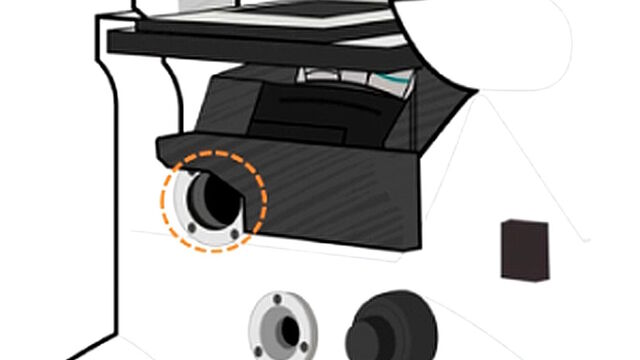
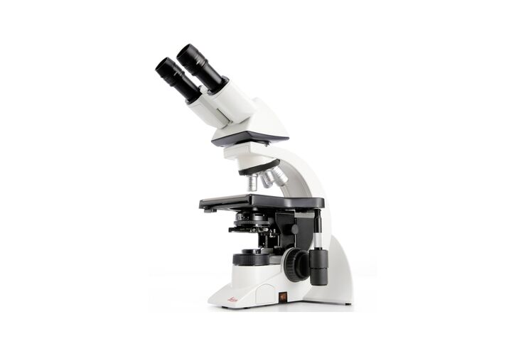
光学顕微鏡
光学顕微鏡
What are you interested in? Show subnavigation
光学顕微鏡
製品紹介 Show subnavigation
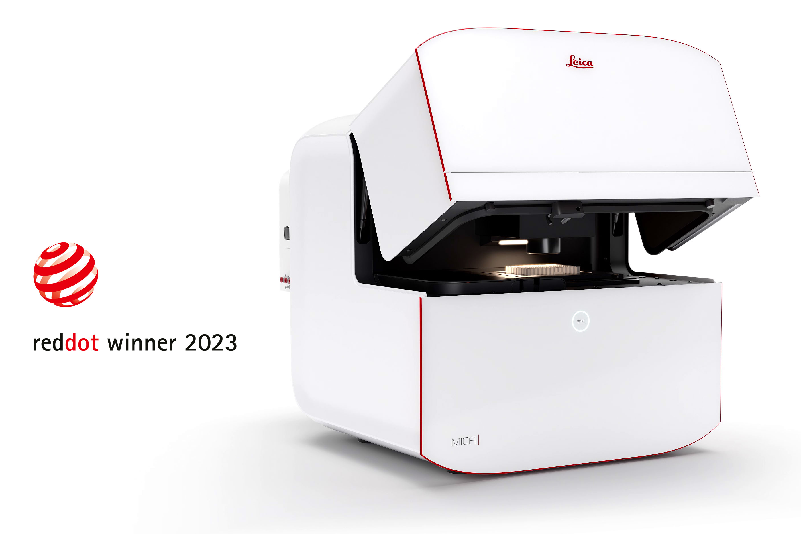
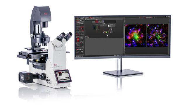
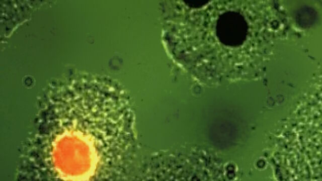
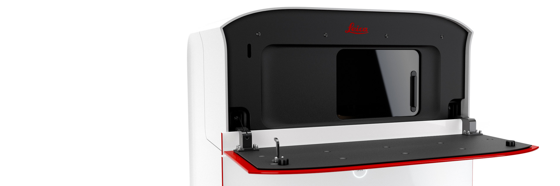
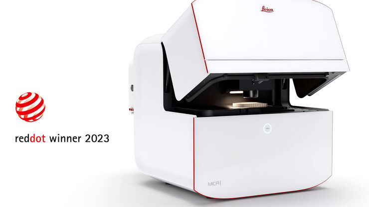
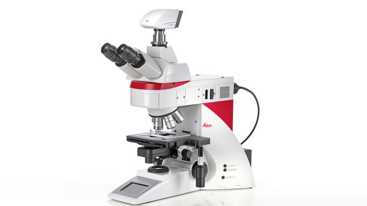
Mica
Mica 発見を可能にするための必要なすべてがひとつの使いやすいシステムに統合 同時 4 色広視野、共焦点分解能、人工知能サポート分析

Visoria P
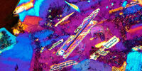
ルーチンの顕微鏡作業も、効率性と快適性の向上を実感いただけます。Visoria P 偏光顕微鏡は、偏光を利用してプラスチックや樹脂、岩石など地質試料の光学特性を観察できます。
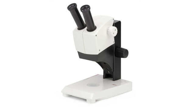
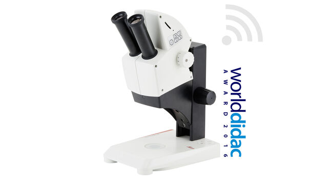
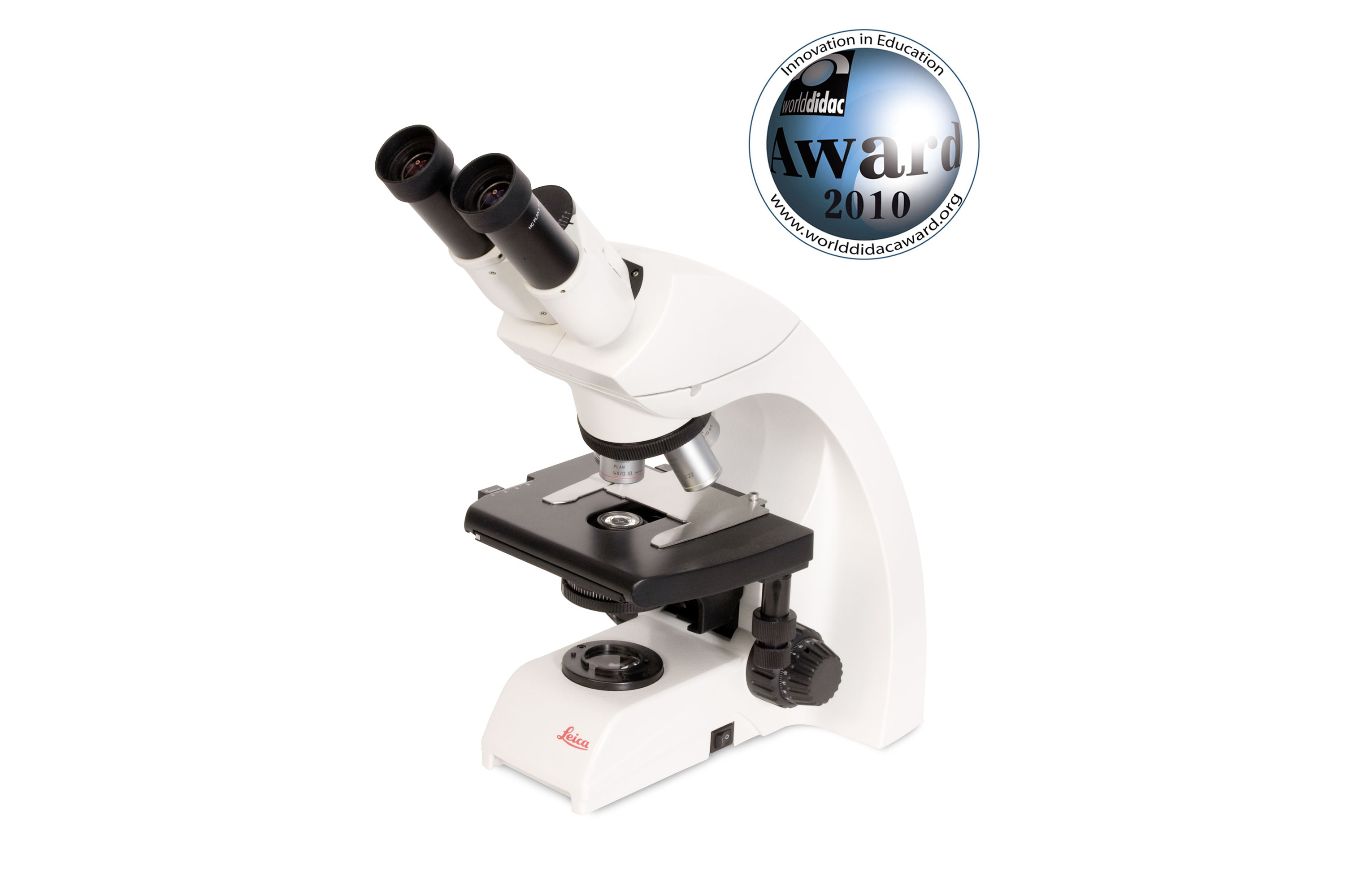
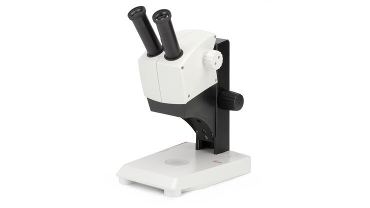
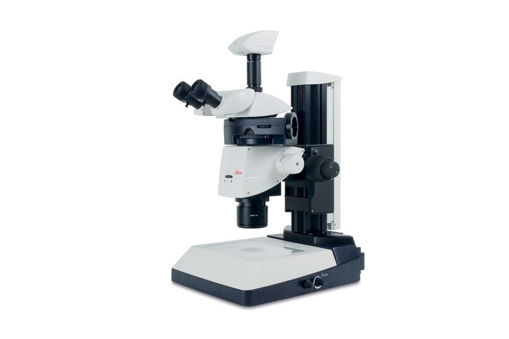
EZ4
立体視可能な実体顕微鏡、8~35倍の拡大観察が可能。最新のLED 照明は7通りの条件設定が可能
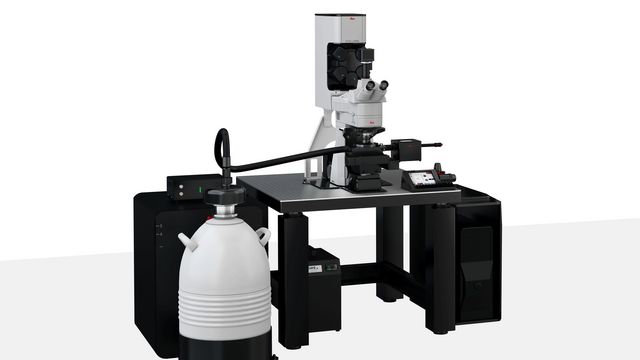
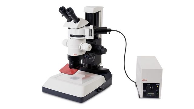
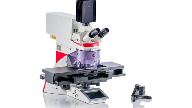
STELLARIS Cryo
STELLARIS Cryoはクライオ電子トモグラフィー (CryoET) の関心領域のターゲティングを効果的に行える共焦点光学顕微鏡です
Leica EZ4 W & EZ4 E
立体視可能な実体顕微鏡、8~35倍の拡大観察が可能。最新のLED 照明は7通りの条件設定が可能。デジタルカメラ内蔵
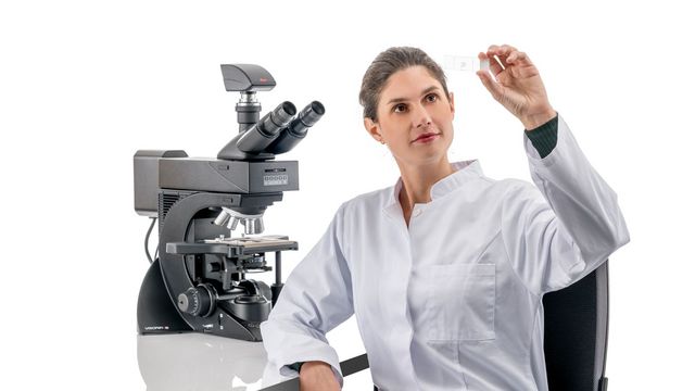
Visoria B
ルーチンの顕微鏡作業も、効率性と快適性の向上を実感いただけます。Visoria B 検査用顕微鏡は、生命科学や臨床検査室で行われる用途向けです。
MZ10 F
ハイコントラストな蛍光観察に実体蛍光顕微鏡
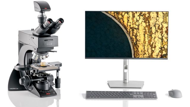
Visoria M
ルーチンの顕微鏡作業も、効率性と快適性の向上を実感いただけます。金属顕微鏡 Visoria Mは、金属、電子、ポリマー業界での用途や、材料科学研究所での用途に適しています。

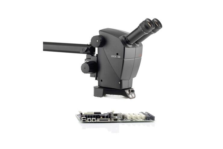
A60 F と A60 S
産業用実体顕微鏡

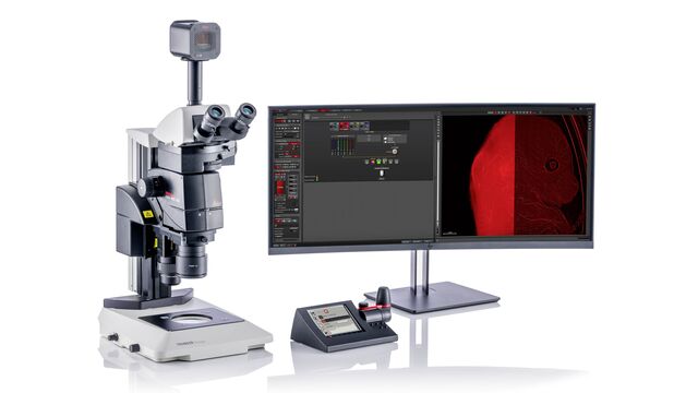
THUNDER Imager Cellスピニングディスク
THUNDER 技術と CrestOptics CICERO スピニングディスクを組み合わせた実力をご覧ください。

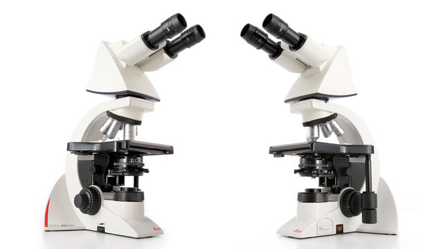
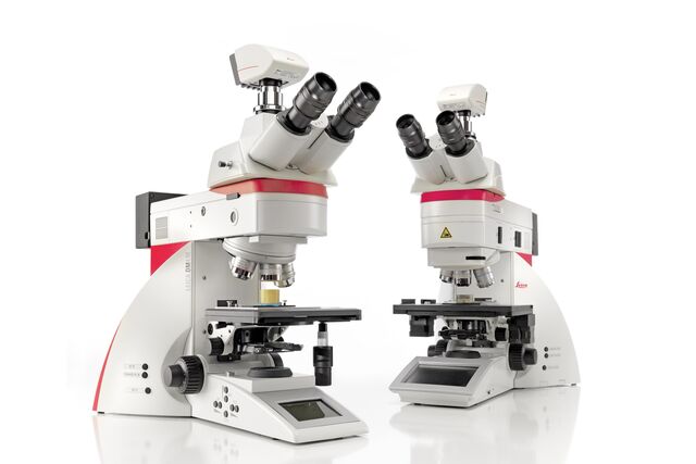
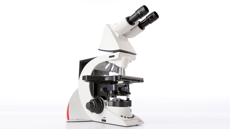
Leica DM3000 & DM3000 LED
インテリジェントなオートメーションを搭載した独自の人間工学に基づいたシステム顕微鏡
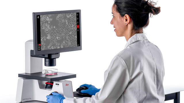
Mateo TL
日常的な細胞チェックと一貫した細胞密度評価を容易にする透過観察用のデジタル顕微鏡
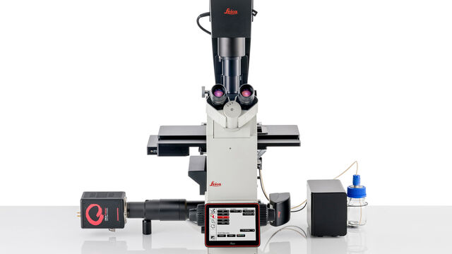
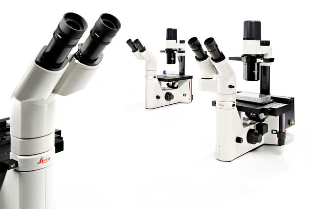
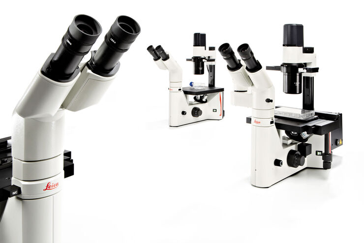
DMi8
DMi8倒立顕微鏡プラットフォームは、お客様の研究条件やご予算に合わせたソリューションで、高品質なデータ取得を可能にします。
DM500
ライフサイエンス用双眼式生物顕微鏡。無限遠補正光学系。4穴レボルバ、蛍光観察も対応可能(オプション)

DM8000 M & DM12000 M
信頼性の高いDM8000 M & DM12000 M検査顕微鏡で、欠陥を検出し、試料の概観を素早く取得できます。
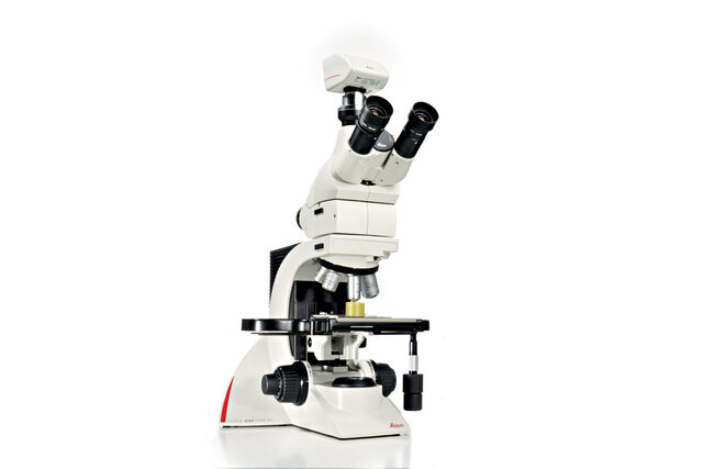
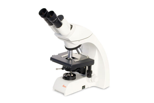
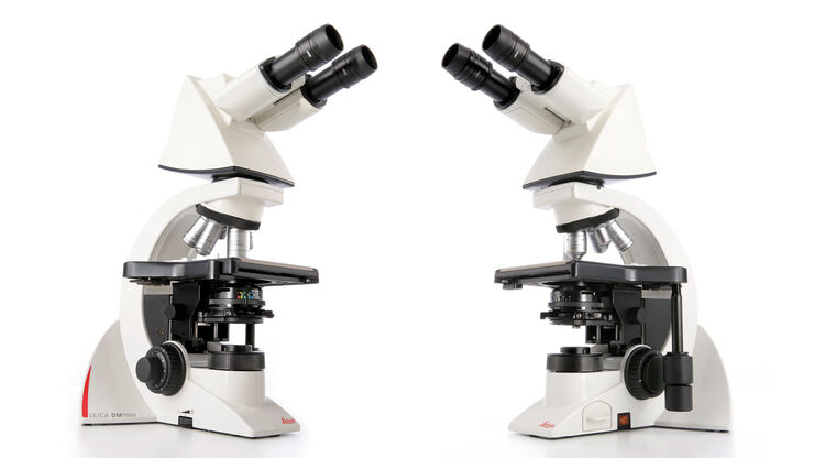
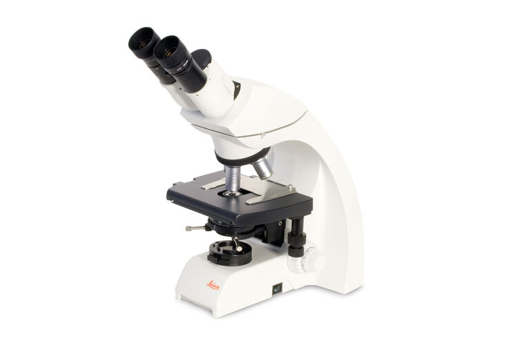
DM1000
ユーザー専用にカスタマイズ
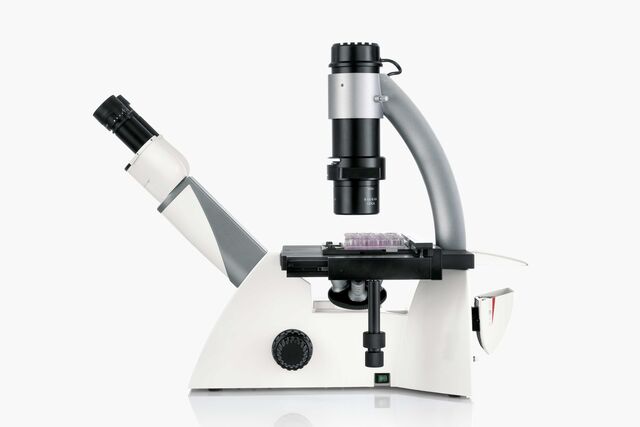
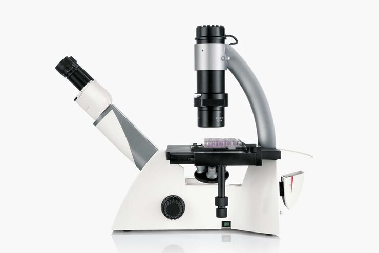
DM IL LED
LED照明を備えた研究用倒立顕微鏡
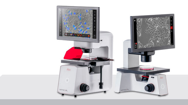
デジタル倒立顕微鏡 Mateo
Mateo FLは、一貫した細胞培養分析のためのAI支援ワークフローを備えた細胞培養研究用の一体型倒立デジタル蛍光顕微鏡です。
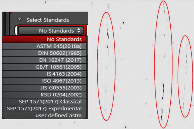
LAS X Steel Expert
非金属介在物の自動解析
Ivesta 3
信頼性の高い結果と共に、目視検査とリワーク作業を最適化します。
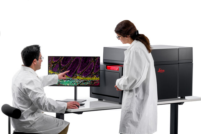
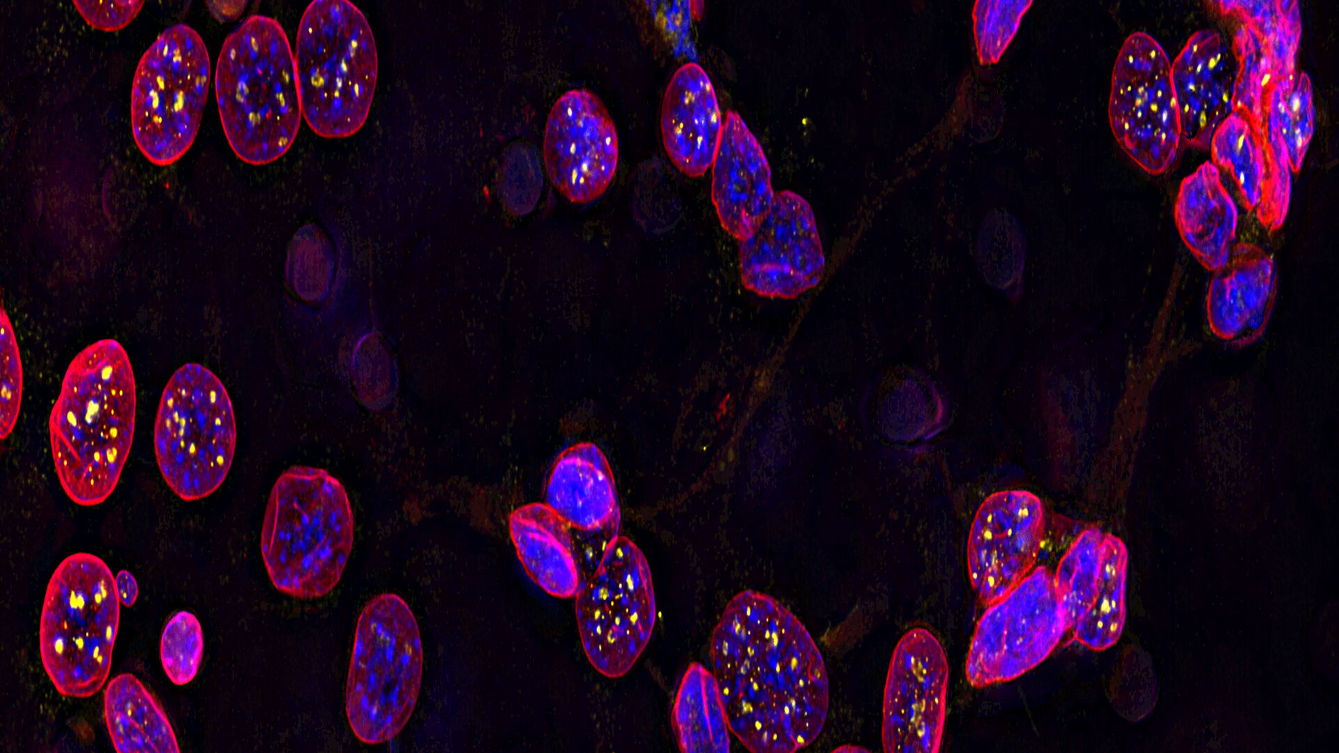
Cell DIVE
マルチプレックスイメージングソリューション「Cell DIVE」は、組織全体の鮮明な画像、60以上のバイオマーカー、350以上の有効な抗体を可視化します。
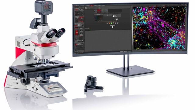
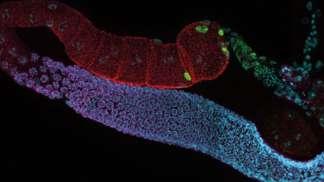
THUNDER モデル生物
THUNDER モデル生物を用いると、発生生物学・分子生物学研究を目的に、素早く簡単に 3D 観察することができます。
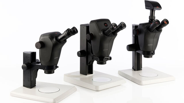
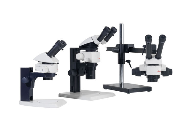
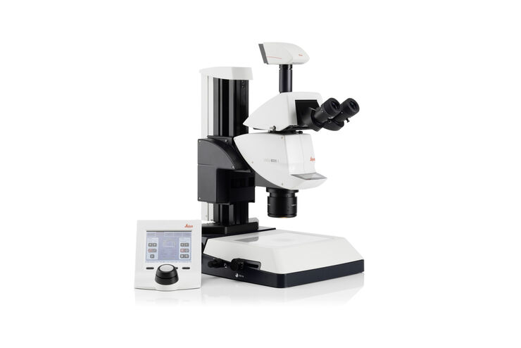
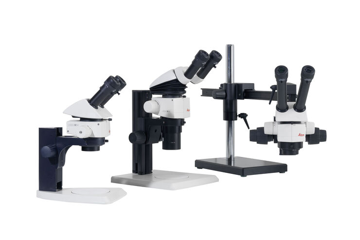
M125 C & M205 C
Mシリーズ実体顕微鏡

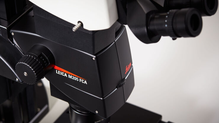
Leica M205 FCA & Leica M205 FA
セミ電動蛍光実体顕微鏡
THUNDER 組織標本
THUNDER 組織標本では、神経科学や組織学研究でよく用いられる組織切片の三次元の蛍光像をリアルタイムに取得することができます。
THUNDER 3D 生細胞および 3D 培養細胞
THUNDER は幹細胞、スフェロイド、オルガノイドなど、高度な 3 次元培養アッセイのためのソリューションです。

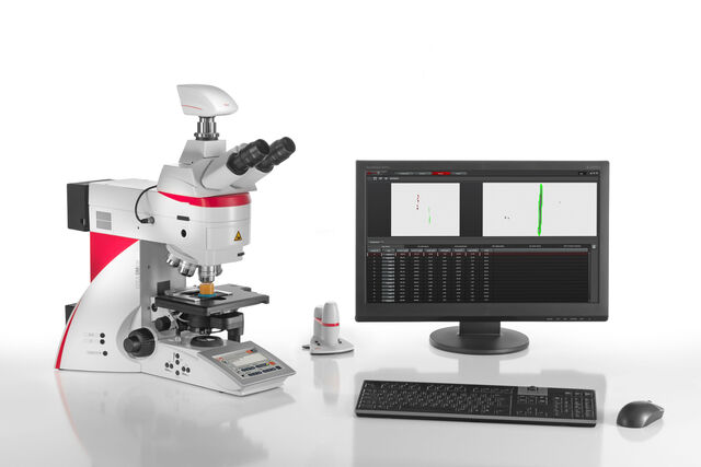
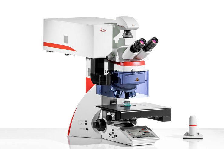
DM6 M LIBS
拡大観察と化学分析を 1 台で
Leica LMD6 & LMD7
レーザーマイクロダイセクション
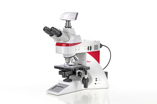
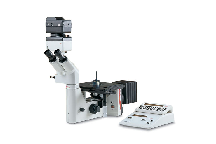
Leica DM4 M & DM6 M
正立金属顕微鏡

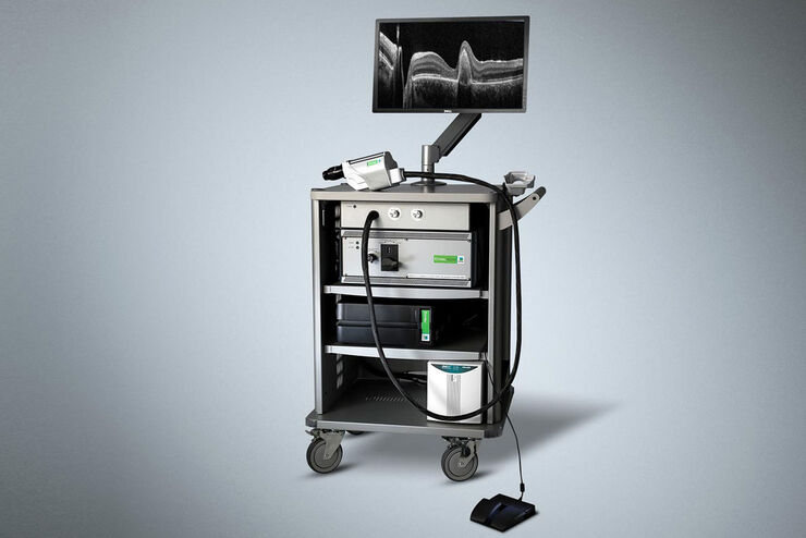
Envisu R-Class
前臨床研究者向け光干渉断層撮影(オプティカル・コヒーレンス・トモグラフィー)
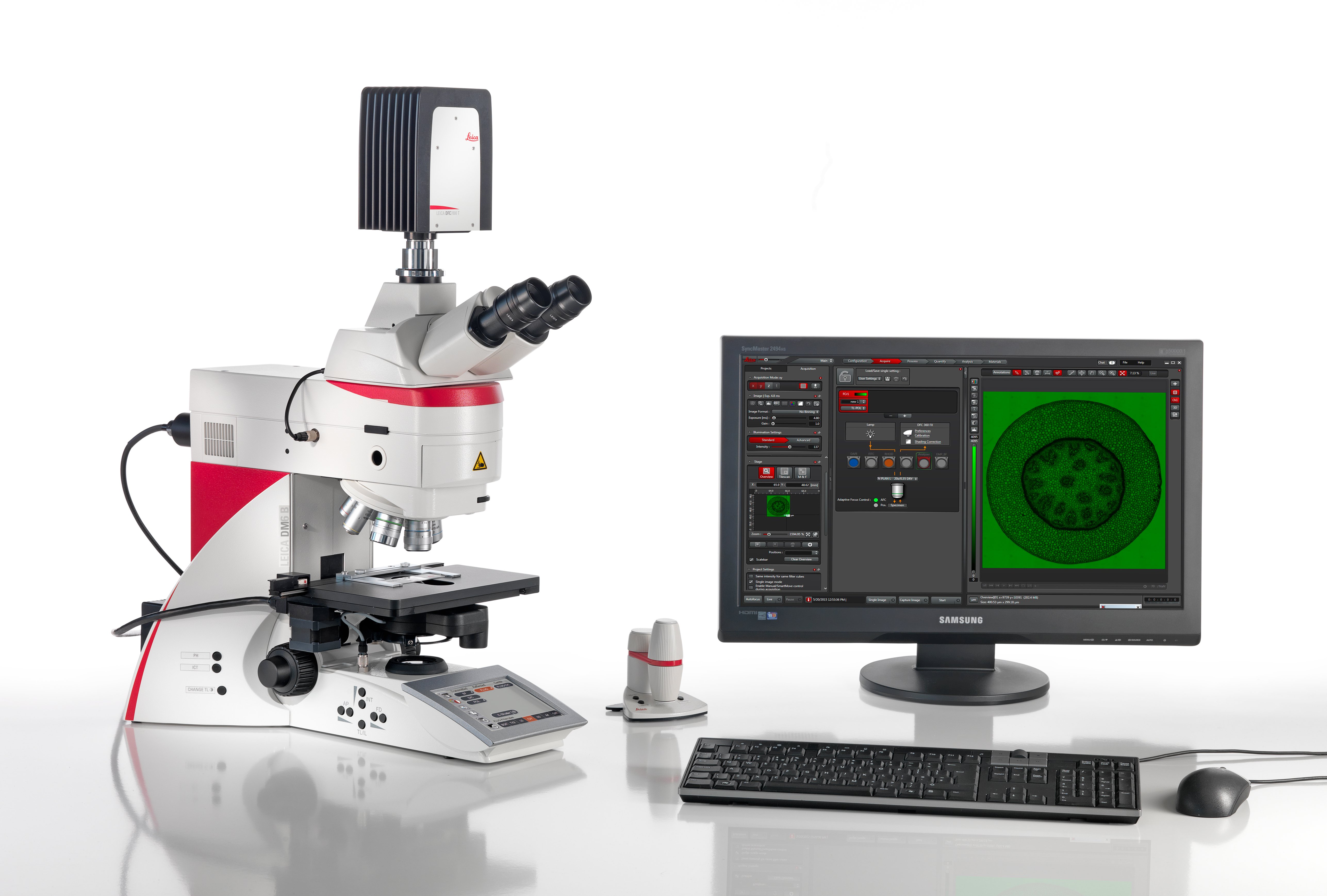
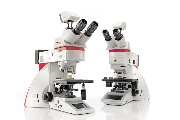
Leica DM4 B & DM6 B
ライフサイエンスと臨床アプリケーションにおけるインテリジェントな自動化

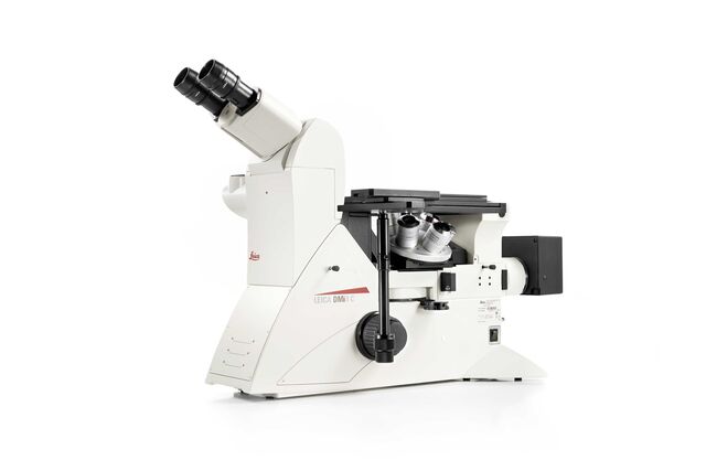
Leica DMi8 M / C / A
工業用倒立顕微鏡
DMi1
Entry level inverted microscope
DM1750 M
材料観察用顕微鏡
M50、M60、M80
ルーチン実体顕微鏡

M165 FC
実体蛍光顕微鏡

DM ILM
倒立金属顕微鏡
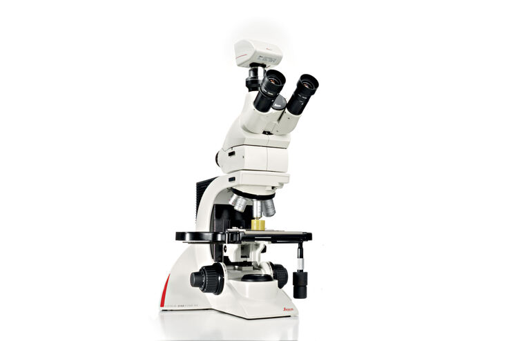
DM750
ライフサイエンス用双眼式生物顕微鏡。無限遠補正光学系。4あるいは5穴レボルバ、観察も対応可能(オプション)

DM1000 LED
検査用顕微鏡
LAS X Widefield Systems
高度な最先端イメージングと解析のための 蛍光顕微鏡システム
Leica Science Lab Show subnavigation
最新の記事を読む
ライカマイクロシステムズのサイエンスラボポータル は、顕微鏡をテーマとする科学研究や記事を提供しています。 コンテンツは、日常業務や実験で、ビギナーから経験豊富な専門家、科学者まで幅広くサポートします。
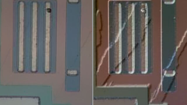
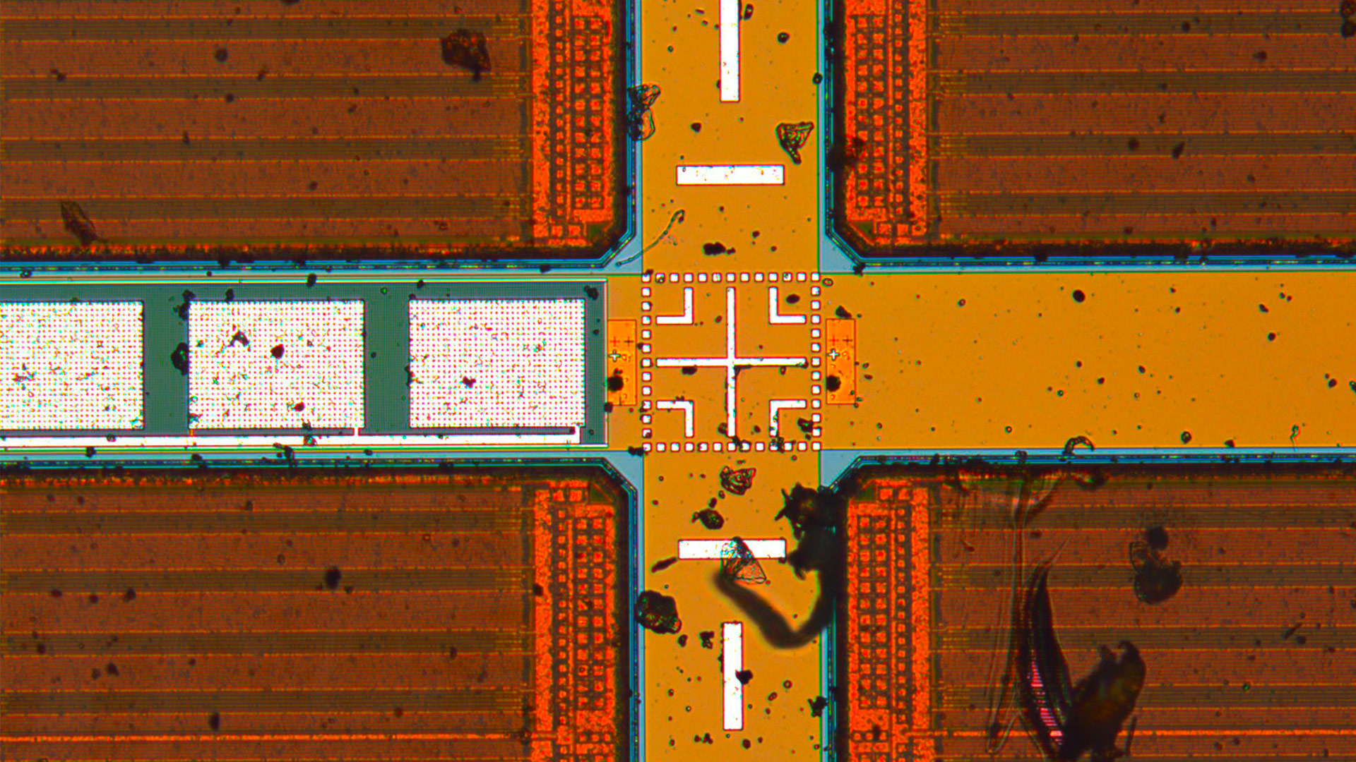
Rapid Semiconductor Inspection with Microscope Contrast Methods
Cross-section Analysis for Electronics Manufacturing
Epi-Illumination Fluorescence and Reflection-Contrast Microscopy
ISO 9022 Standard Part 11 - Testing Microscopes with Severe Conditions
Life Science Research: Which Microscope Camera is Right for You?
Factors to Consider When Selecting a Research Microscope

Challenges Faced When Manually Rating Non-Metallic Inclusions (NMIs) to Determine Steel Quality
Top Issues Related to Standards for Rating Non-Metallic Inclusions in Steel
Analyzing Non-metallic Inclusions in Steel
Rate the Quality of Your Steel: Free Webinar and Report
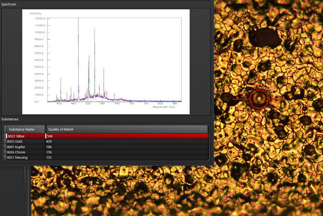
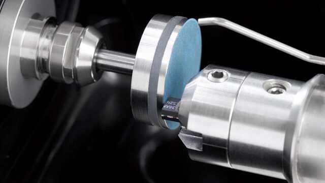
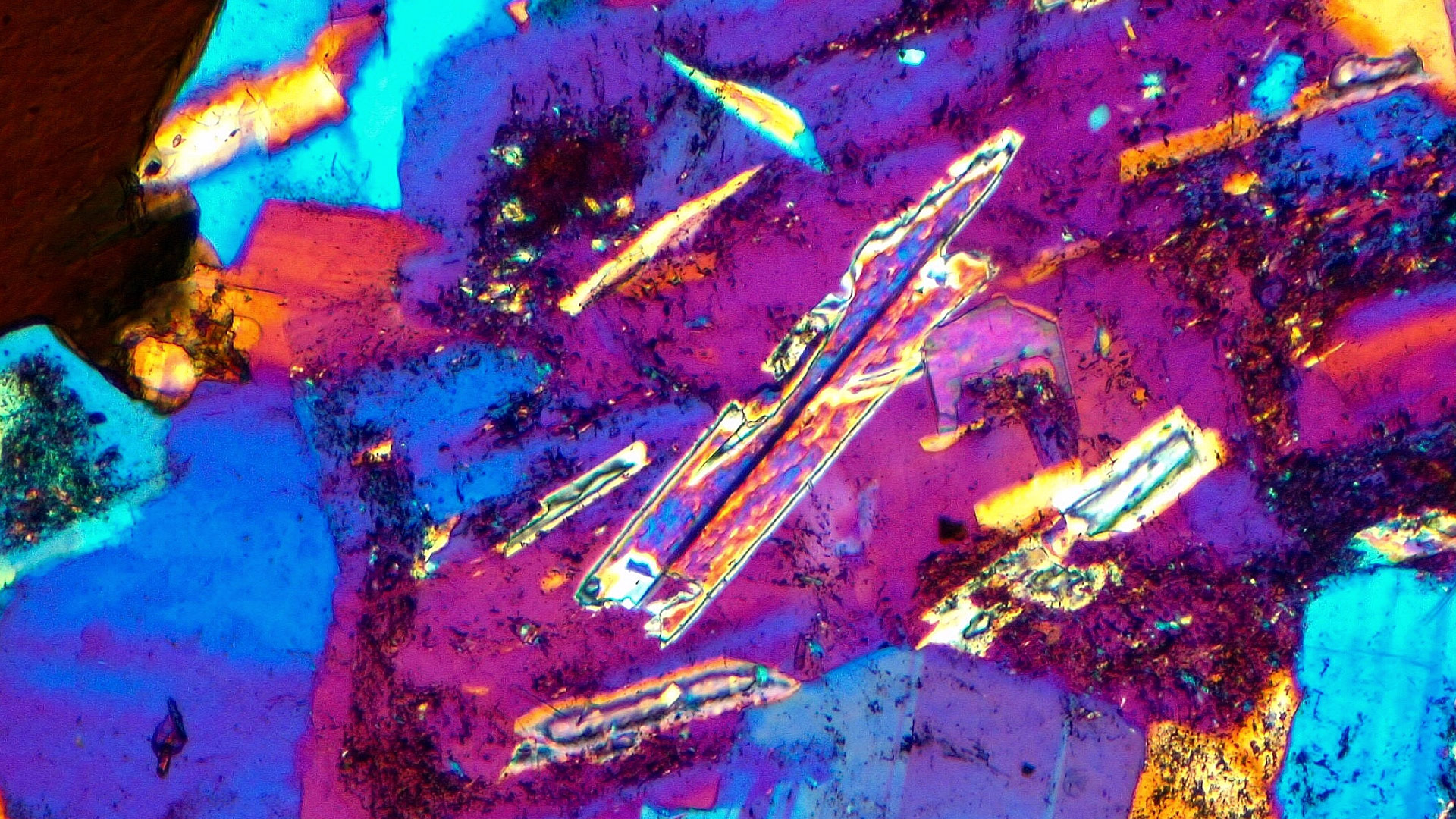
See the Structure with Microscopy - Know the Composition with Laser Spectroscopy
Optimization of the Interplay of Optical Components for Aberration Free Microscopy
Infinity Optical Systems
Key Questions
1主な用途は何ですか?
ライカの複合光学顕微鏡のうち、どれが最適となるかは、用途によって異なります。 様々な用途におけるニーズを満たすには、多様な種類の光学顕微鏡と光学顕微鏡パーツを用いることが最善の方法です。 ライカの光学顕微鏡は、モジュール設計を採用しており、特定用途に合わせたカスタマイズが可能です。
2どのような種類の試料を可視化する必要がありますか?
ライカの光学顕微鏡ソリューションは、日常的なラボ作業、素材の生産や分析、複雑なライフサイエンス研究など、試料の観察に必要な光学解像度、コントラスト、被写界深度、画質を提供します。 さらに、対物レンズ、照明タイプ、デジタルカメラなど、光学顕微鏡のパーツやアクセサリー、Leica Application Suite X(LAS X) ソフトウェアにより、お客様の具体的な用途におけるニーズに合わせたソリューションのカスタマイズや最適化を行うことができます。
3倒立顕微鏡ソリューションの予算は?
カスタマイズされた光学顕微鏡ソリューションは、投資コストが高くなる可能性はありますが、チームの生産性を高めることができます。 光学顕微鏡、光学顕微鏡部品、アクセサリーの豊富なバリエーションにより、あらゆる用途において、最適な顕微鏡ソリューションが実現します。
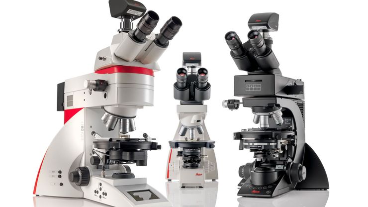
4複合光学顕微鏡と実体顕微鏡の違いは何ですか?
複合光学顕微鏡と実体顕微鏡は、対物レンズと接眼レンズを備えた光学系を使用して、試料を観察します 一般的に、複合光学顕微鏡は実体顕微鏡よりも幅広い倍率に対応します。 複合顕微鏡では、2次元の像を観察できます。 実体顕微鏡は、観察者の左右の目で異なる光路を持ち、試料を立体視することができます。 複合顕微鏡や実体顕微鏡にカメラが搭載されている場合は、光路を1つ使用しますので、2次元の画像しか記録することができません。
光学顕微鏡 41
Mica

Visoria P
EZ4
光学顕微鏡
Filter by Area of Application
Mica
Visoria P
EZ4

STELLARIS Cryo

Leica EZ4 W & EZ4 E

Visoria B

MZ10 F

Visoria M
A60 F と A60 S

THUNDER Imager Cellスピニングディスク
Leica DM3000 & DM3000 LED

Mateo TL

DMi8

DM500

DM8000 M & DM12000 M
DM1000
DM IL LED

デジタル倒立顕微鏡 Mateo

LAS X Steel Expert

Ivesta 3

Cell DIVE

THUNDER モデル生物
M125 C & M205 C
Leica M205 FCA & Leica M205 FA

THUNDER 組織標本

THUNDER 3D 生細胞および 3D 培養細胞
DM6 M LIBS

Leica LMD6 & LMD7
Leica DM4 M & DM6 M
Envisu R-Class
Leica DM4 B & DM6 B
Visoria P、DM750 P、DM4 P

Leica DMi8 M / C / A
DMi1
DM1750 M
M50、M60、M80
M165 FC
DM ILM
DM750
DM1000 LED

LAS X Widefield Systems
もっと知りたいですか?
お気軽にお問合せください
ライカまでお気軽にご相談ください Show local contacts
Leica Microsystems Inc.
フォームを閉じてよろしいですか?
no yes
