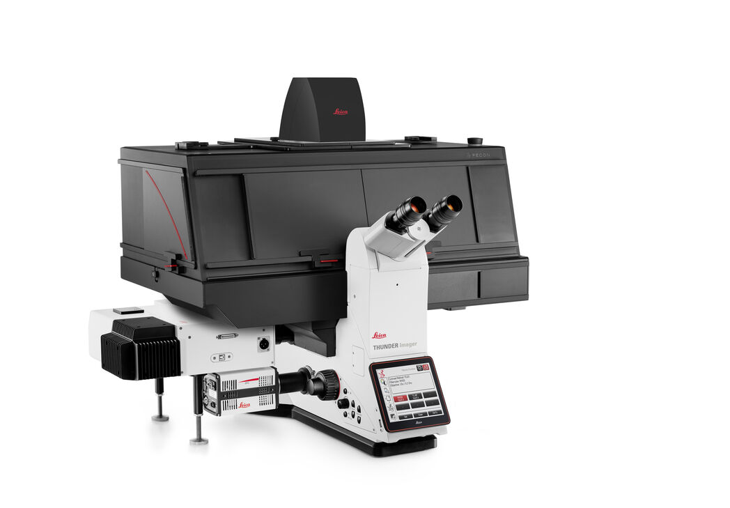
DMi8 S
Inverted
Light Microscopes
Products
Home
Leica Microsystems
DMi8 S Inverted Microscope Solution
Find the missing links
Read our latest articles
Differential Interference Contrast (DIC) Microscopy
This article demonstrates how differential interference contrast (DIC) can be actually better than brightfield illumination when using microscopy to image unstained biological specimens.
Phase Contrast and Microscopy
This article explains phase contrast, an optical microscopy technique, which reveals fine details of unstained, transparent specimens that are difficult to see with common brightfield illumination.
Immersion Objectives
How an immersion objective, which has a liquid medium between it and the specimen being observed, helps increase the numerical aperture and microscope resolution is explained in this article.
Precise Spatial Proteomic Information in Tissues
Despite the availability of imaging-based and mass-spectrometry-based methods for spatial proteomics, a key challenge remains connecting images with single-cell-resolution protein abundance…
Studying Wound Healing of Smooth Muscle Cells
This article discusses how wound healing of cultured smooth muscle cells (SMCs) in multiwell plates can be reliably studied over time with less effort using a specially configured Leica inverted…
How to Prepare your Specimen for Immunofluorescence Microscopy
Immunofluorescence (IF) is a powerful method for visualizing intracellular processes, conditions and structures. IF preparations can be analyzed by various microscopy techniques (e.g. CLSM,…
Fluorescent Dyes
A basic principle in fluorescence microscopy is the highly specific visualization of cellular components with the help of a fluorescent agent. This can be a fluorescent protein – for example GFP –…
Studying Actin Cytoskeleton and Microfilament Assembly
This article demonstrates how actin microfilament assemblies can be clearly visualized with superior resolution at the plasma membrane of a cell using a DMi8 S Infinity TIRF High Power imaging…
Download The Guide to Live Cell Imaging
In life science research, live cell imaging is an indispensable tool to visualize cells in a state as in vivo as possible. This E-book reviews a wide range of important considerations to take to…
Plant Cell Development and Morphogenesis
The article discusses how tubulin molecules, which make up microtubules, in plant cells can be studied with total internal reflection fluorescence (TIRF) microscopy to better understand tubulin…
Factors to Consider When Selecting a Research Microscope
An optical microscope is often one of the central devices in a life-science research lab. It can be used for various applications which shed light on many scientific questions. Thereby the…
Upright Fluorescence Microscopy for Virus Replication Studies
The severe acute respiratory syndrome coronavirus 2 (SARS-CoV-2) emerged in late 2019 and spread over the world quickly. Due to its dramatic impact, researchers are interested in the nature of the…
Microscopy in Virology
The coronavirus SARS-CoV-2, causing the Covid-19 disease effects our world in all aspects. Research to find immunization and treatment methods, in other words to fight this virus, gained highest…
Where to go? Cellular Migration requires coordinated Transitions of Actin Cortex
Plants, Bacteria, and Fungi possess a rigid cell wall that protects the cell and gives it shape. Animal cells, such as mammalian cells, have no outer wall, which exposes their plasma membrane to the…
Koehler Illumination: A Brief History and a Practical Set Up in Five Easy Steps
The technique of Koehler Illumination is one of the most important and fundamental techniques in achieving optimum imaging in any given light microscope set-up. Although it should be routinely used as…
Introduction to Mammalian Cell Culture
Mammalian cell culture is one of the basic pillars of life sciences. Without the ability to grow cells in the lab, the fast progress in disciplines like cell biology, immunology, or cancer research…
Introduction to Widefield Microscopy
This article gives an introduction to widefield microscopy, one of the most basic and commonly used microscopy techniques. It also shows the basic differences between widefield and confocal…
What is Photomanipulation?
The term photomanipulation describes a wide range of techniques that enable the microscopist the transition from passive observer to instigator of events by offering a way of interacting with their…
Photoactivatable, Photoconvertible, and Photoswitchable Fluorescent Proteins
Fluorescent proteins (FPs) such as GFP, YFP or DsRed are powerful tools to visualize cellular components in living cells. Nevertheless, there are circumstances when classical FPs reach their limits.…
Milestones in Incident Light Fluorescence Microscopy
Since the middle of the last century, fluorescence microscopy developed into a bio scientific tool with one of the biggest impacts on our understanding of life. Watching cells and proteins with the…
Chronic Inflammation Under the Microscope
In the course of chronic inflammation certain body areas are recurrently inflamed. This goes along with many human diseases. With the help of widefield light microscopy, the underlying processes can…
Gene Editing with CRISPR/Cas9 - Breakthrough in Genome Engineering
The CRISPR/Cas9 system is one of several different bacterial systems for defense against viral attacks. It consists of two main components. One is a small piece of RNA which binds to the viral target…
Work Efficiently in Developmental Biology with Stereo and Confocal Microscopy: C. elegans
For scientists, technicians, and teachers working with the worm C. elegans in the research lab or classroom, this report is intended to give useful information to help improve their daly work. The aim…
Infinity Optical Systems
“Infinity Optics” refers to the concept of a beam path with parallel rays between the objective and the tube lens of a microscope. Flat optical components can be brought into this “Infinity Space”…
Video Talk by Daniel Axelrod: Total Internal Reflection Fluorescence (TIRF) Microscopy
Total Internal Reflection Fluorescence (TIRF) Microscopy is a technique that only illuminates dye molecules near a surface. In this video, the pioneer of TIRF Microscopy describes what this technique…
Handbook of Optical Filters for Fluorescence Microscopy
Fluorescence microscopy and other light-based applications require optical filters that have demanding spectral and physical characteristics. Often, these characteristics are application-specific and…
Optogenetics
Optogenetics is a technique that allows light-controlled responses of transfected cells. The cells are genetically modified by introduction of genes that code for light-induced channels or ion pumps.…
Video Tutorial: How to Align the Bulb of a Fluorescence Lamp Housing
The traditional light source for fluorescence excitation is a fluorescence lamp housing with mercury burner. A prerequisite for achieving bright and homogeneous excitation is the correct centering and…
Video Tutorial: How to Change the Bulb of a Fluorescence Lamp Housing
When applying fluorescence microscopy in biological applications, a lamp housing with mercury burner is the most common light source. This video tutorial shows how to change the bulb of a traditional…
Controlling the TIRF Penetration Depth is Mandatory for Reproducible Results
The main feature of total internal reflection fluorescence (TIRF) microscopy is the employment of an evanescent wave for the excitation of fluorophores instead of using direct light. A property of the…
Basic Principles of Luminescence
There are a lot of light-emitting processes occurring in nature. Luminescence is an umbrella term for those kinds of events where light emission is not the result of high temperatures. This article…
Fluorescent Proteins – Introduction and Photo Spectral Characteristics
The prospects of fluorescence microscopy changed dramatically with the discovery of fluorescent proteins in the 1950s. The starting point was the detection of the jellyfish Aequorea victoria green…
Total Internal Reflection Fluorescence (TIRF) Microscopy
Total internal reflection fluorescence (TIRF) is a special technique in fluorescence microscopy developed by Daniel Axelrod at the University of Michigan, Ann Arbor in the early 1980s. TIRF microscopy…
Fluorescent Proteins - From the Beginnings to the Nobel Prize
Fluorescent proteins are the fundament of recent fluorescence microscopy and its modern applications. Their discovery and consequent development was one of the most exciting innovations for life…
Fluorescence Recovery after Photobleaching (FRAP) and its Offspring
FRAP (Fluorescence recovery after photobleaching) can be used to study cellular protein dynamics: For visualization the protein of interest is fused to a fluorescent protein or a fluorescent dye. A…
Optical Contrast Methods
Optical contrast methods give the potential to easily examine living and colorless specimens. Different microscopic techniques aim to change phase shifts caused by the interaction of light with the…
Integrated Modulation Contrast (IMC)
Hoffman modulation contrast has established itself as a standard for the observation of unstained, low-contrast biological specimens. The integration of the modulator in the beam path of themodern…
An Introduction to Fluorescence
Fluorescence is widely used in microscopy and an important tool for observing the distribution of specific molecules. Most molecules in cells do not fluoresce. They therefore have to be marked with…
Fluorescence in Microscopy
Fluorescence microscopy is a special form of light microscopy. It uses the ability of fluorochromes to emit light after being excited with light of a certain wavelength. Proteins of interest can be…
TIRF Microscopy of the Apical Membrane of Polarized Epithelial Cells
Application of TIRF microscopy (Total Internal Reflection Fluorescence) allows the visualization of structures at the apical surface of polarized epithelial cells that have been hidden in conventional…
Exploring Cell Logistics
Using TIRF microscopy, scientists have been able to take a closer look at intracellular transport processes with the example of the galactose-binding protein Galectin-3, which has been identified as a…
Multi-Wavelength Epi-Illumination in Fluorescence Microscopy
Fluorescence is a process where a substance after having absorbed
light (photons) emitts a radiation the wavelength (colour) of which is
longer than that of the absorbed light, and where this…
Fields of Application
Fluorescence
Find out how fluorescence microscopes from Leica Microsystems support your research. Fluorescence is one of the most commonly used physical phenomena in biological and analytical microscopy for its…
Micromanipulation
Witness automated, fast micromanipulation with this easy and convenient system from Leica Microsystems. Fully equipped with inverted research microscope and electronic micromanipulators, these…
Photomanipulation
The term photomanipulation encompasses a range of techniques that utilize the properties of fluorescent molecules to initiate events and observe how dynamic complexes behave over time in living cells.…
Live Cell Imaging
Shifting perspective from single microscope components to a full working live cell imaging solution, Leica Microsystems integrates microscope, LAS X imaging software, cameras, and dedicated…
Cell Culture
Growing cells under lab conditions is the base for scientists working in fields of cell or developmental biology, cancer research, or any kind of life science and pharma research. Find out how Leica…
Virology
Do your research interests focus on viral infection and disease? Find out how you can gain insights into virology with solutions for imaging and sample preparation from Leica Microsystems.
Advanced Microscopy Techniques
Advanced microscopy techniques: Advanced microscopy techniques encompass both high-resolution and super-resolution imaging techniques. These techniques are primarily used to visualize biological…
Basic Microscopy Techniques
Basic microscopy techniques are used in instances where the entire specimen on the microscope stage is exposed to a light source. The whole specimen is illuminated by white light either from above (in…
Darkfield Microscopes
The darkfield contrast method exploits diffraction or scattering of light from structures of a biological specimen or non-uniform features of a material sample.
Phase Contrast Light Microscopes
A phase contrast light microscope offers a way to view the structures of many types of biological specimens in greater contrast without the need of stains.
DIC Microscopes
A DIC microscope is a widefield microscopy which has a polarization filter and Wollaston prism between the light source and condenser lens and also between the objective lens and camera sensor or…