Contact Us

K3C & K3M The Microscope Camera K3 series

For Life Science and Industry Imaging Applications and Analysis

Read our latest articles

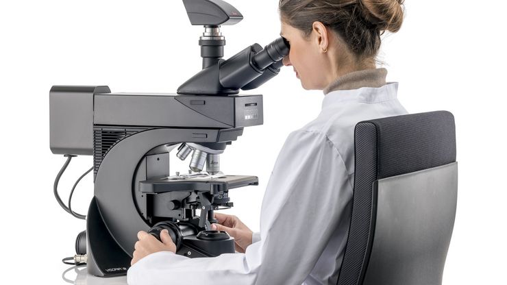
Microscope equipped with a K7 color CMOS camera for life-science and industry imaging applications.

Technical Terms for Digital Microscope Cameras and Image Analysis

Learn more about the basic principles behind digital microscope camera technologies, how digital cameras work, and take advantage of a reference list of technical terms from this article.
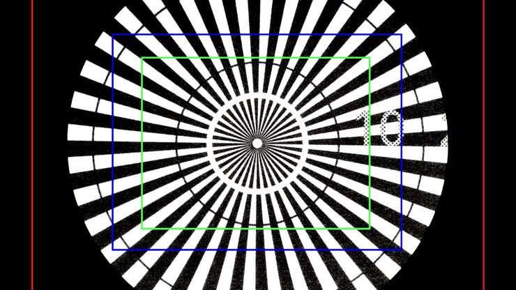
Image of a Siemens star, where the diameter of the 1st black line circle is 10 mm and the 2nd is 20 mm, taken via an eyepiece of a M205 A stereo microscope. The rectangles represent the field of view (FOV) of a Leica digital camera when installed with various C-mounts (red 0.32x, blue 0.5x, green 0.63x).

Understanding Clearly the Magnification of Microscopy

To help users better understand the magnification of microscopy and how to determine the useful range of magnification values for digital microscopes, this article provides helpful guidelines.
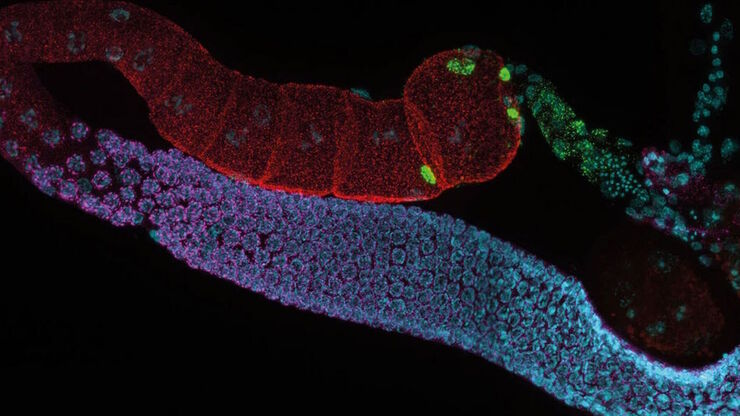
C. elegans adult hermaphrodite gonades acquired using THUNDER Imager. Staining: blue - DAPI (nucleus), green - SP56 (sperm), red - RME-2 (oocyte), magenta - PGL-1 (RNA + protein granules). Image courtesy of Prof. Dr. Christian Eckmann, Martin Luther University, Halle, Germany.

Life Science Research: Which Microscope Camera is Right for You?

Deciding which microscope camera best fits your experimental needs can be daunting. This guide presents the key factors to consider when selecting the right camera for your life science research.
Laboratory

Factors to consider when selecting clinical microscopes

What matters if you would like to purchase a clinical microscope? Learn how to arrive at the best buying decision from our Science Lab Article.
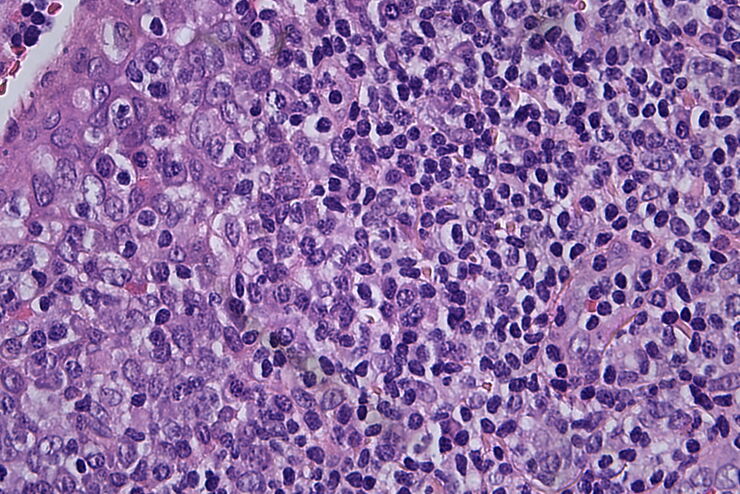
Histopathological sample, 40x magnification

Clinical Microscopy: Considerations on Camera Selection

The need for images in pathology laboratories has significantly increased over the past few years, be it in histopathology, cytology, hematology, clinical microbiology, or other applications. They…
H&E stained specimen, 20x magnification

The Time to Diagnosis is Crucial in Clinical Pathology

Abnormalities in tissues and fluids - that’s what pathologists are looking for when they examine specimens under the microscope. What they see and deduce from their findings is highly influential, as…

Fields of Application

Cancer Research

Cancer is a complex and heterogeneous disease caused by cells deficient in growth regulation. Genetic and epigenetic changes in one or a group of cells disrupt normal function and result in…

Microscopy Solutions for Cell Culture

Growing cells under lab conditions is the base for scientists working in fields of cell or developmental biology, cancer research, or any kind of life science and pharma research. Find out how Leica…

Fluorescence

Find out how fluorescence microscopes from Leica Microsystems support your research. Fluorescence is one of the most commonly used physical phenomena in biological and analytical microscopy for its…

Laser Capture Microdissection

Laser Microdissection, also known as LMD or LCM (Laser Capture Microdissection), is a contact- and contamination-free method for isolating specific single cells or entire areas of tissue from a wide…

Micromanipulation

Witness automated, fast micromanipulation with this easy and convenient system from Leica Microsystems. Fully equipped with inverted research microscope and electronic micromanipulators, these…

Semiconductor Inspection

Achieve fast and reliable wafer and semiconductor inspection for wafer processing, as well as IC packaging, assembly, and testing, with microscope and sample preparation solutions.

PCB Inspection

For rapid and reliable PCB inspection, electronics manufacturers and suppliers require solutions which enable efficient QC, cross-section analysis, and R&D.

Energy, Mining and Natural Resources

Energizing the worlds infrastructure while maintaining sustainability is always a priority. Leica Microsystems has powerful and customizable microscopy solutions to help you get the most out of your…

Forensic Science

As a forensic scientist, your microscopes and imaging equipment must provide precision, quality, accuracy, and reproducibility of results to ensure success when examining evidence. Leica Microsystems…

Clinical Pathology

Learn how ergonomic microscopes from Leica Microsystems support accurate and timely diagnosis in clinical pathology.

Microscopy in Pathology

Analysis of specimens for pathology sometimes requires long hours working with a microscope. The result for the user may be physical discomfort and strain that can lead to reduced efficiency and the…
Background image
Scroll to top