Not all products or services are approved or offered in every market, and approved labelling and instructions may vary between countries. Please contact your local representative for further information.
The THUNDER Imager Model Organism allows fast and easy 3D exploration of whole organisms for developmental or molecular biology research. Thanks to THUNDER Live, your images reveal the finest structural details already in the live image. No hassle with out-of-focus blur while maintaining the capabilities and ease-of-use typical for Leica stereo microscopes.
A THUNDER Imager Model Organism is the optimal instrument for studying, e.g., Drosophila, C. elegans, zebrafish, plants, and mice. One device for screening, positioning, and imaging your specimen. Simplify your workflow and study model organisms from a large overview to the highest detail.
Get the most information from your precious model organisms and access stunning details. Take advantage of the leap forward in image quality with THUNDER Imager Model Organism compared to conventional stereo microscopes. Thanks to THUNDER, non-relevant background is removed and interesting details are preserved.
Obtain results faster and with higher significance for applications like:
Characterization of model organism transgenic lines
Detailed observation of model organisms in real time
Investigating development of neuronal networks
THUNDER Imager Model Organsim with 1x Plan APO objective, zoom factor 11:1, 18 z-slice stack, circa 300 um z-depth. Green (kdrl:GFP) shows angiogenisis and red (GATA1:dsRed) shows red blood cells and platelets.
Image courtesy: Dr. Almary Guerra & Dr. Didier Stainier, Max Planck Institute for Heart and Lung Research, Bad Nauheim (Germany)
Image large model organisms under excellent physiological conditions
Have the best of both worlds: THUNDER Imager Model Organism combines a large field of view with high image quality for thick samples that was, so far, only possible with optical sectioning methods. This powerful combination allows you to observe large specimens live under excellent physiological conditions. No need to sedate your organisms.
Gain more insights for a better understanding of the phenomena under investigation. Z-scans through the entire volume of a fish are possible thanks to the excellent optics and removal of out-of-focus signals. Get fast results as just one image per z-plane is required.
With THUNDER Imager Model Organism, you can observe the relevant details in the proper context with a large field of view. With instant Computational Clearing, you can acquire fast z-stack images and use highly sensitive cameras for detection under physiological conditions.
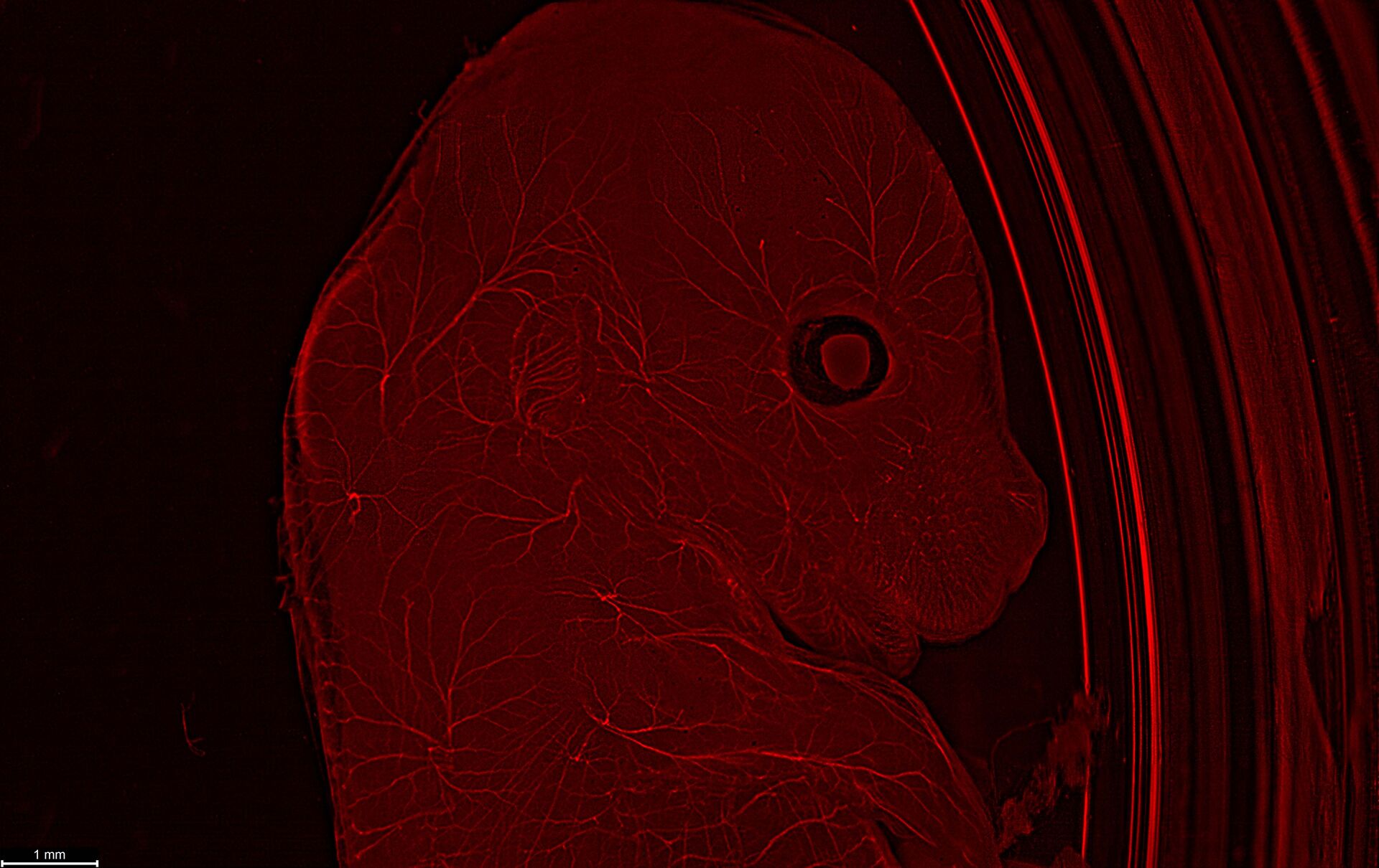
In this E12-14 mouse (wt sample), neurofilaments are stained in red to assess neuronal outgrowth. The mouse was uncleared. Courtesy of Yves Lutz, Centre d’imagerie, IGBMC, France.
With the help of Instant Computational Clearing, I can already localize and evaluate certain signals in brain regions.
The THUNDER Imager Model Organism helps to simplify the screening and documentation of live model organisms for specific details. Live, moving organisms can be imaged directly in petri dishes or multiwell plates. With the optional 2x corr objective, you can observe specimens in water with sharp focus. There is no need to extract or cut the specimen into sections. Just obtain the relevant data directly.
Time-lapse studies of model organisms are important for looking at development, movement, and reaction. With THUNDER, you obtain images with highly resolved details at the speed of your camera, in real time.
THUNDER Imager for deep insights
Take advantage of the large working distances typical for stereo microscopes
THUNDER Imager Model Organism can make z-stack images, which was not possible before. Look at large organoids and cleared or uncleared mouse organs directly in the petri dish. Your results will be highly valuable and simply impressive.
Key Advantages of the THUNDER Imager Model Organism
Our product managers talk about key advantages that the THUNDER Imager Model Organism offer.

 - THUNDER Imager Model Organism")