")
First magnifying instrument built by a spectacle maker
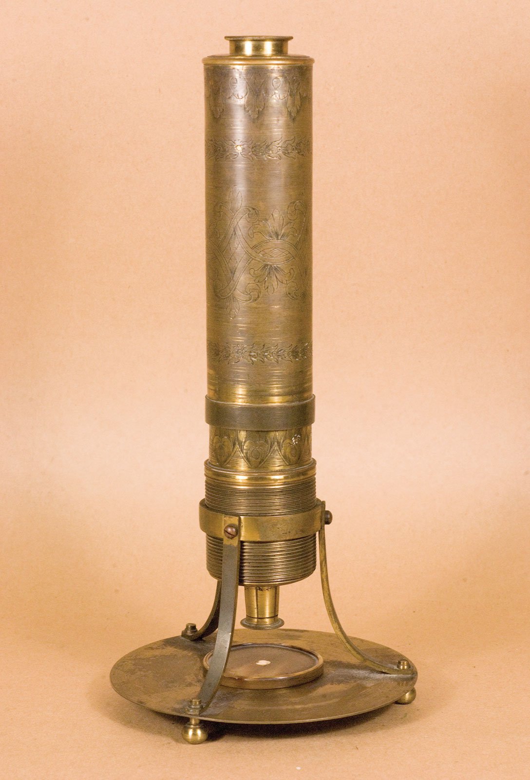
Although the idea of magnifying objects with two glass lenses positioned one in front of the other originated as early as the beginning of the 16th century, it was some time before such an instrument was built. The Dutch spectacle maker Hans Janssen and his son Zacharias are generally credited with creating these compound microscopes. The two of them built what was probably the first compound microscope in the last decade of the 16th century. It had a magnification that could be adjusted between 3 and 9x.
But other scientists applied themselves to building these magnifying instruments, too. In 1609, Galileo Galilei made a microscope by converting one of his telescopes. It had a diverging lens as an eyepiece and a converging lens as an objective. An early microscope made of two converging lenses was presented around 1620 by the astronomer Cornelius Drebbel. However, it was apparently not Drebbel’s own idea but that of Johannes Kepler.


Hooke’s Micrographia
It was the English universal scholar Robert Hooke who really put the relatively new science of microscopy on the map.
In 1667 he was the first to publish a fundamental work on the subject called "Micrographia". The drawings it contained of his observations with the microscope made the microcosm accessible to a wider public. Some of the pictures he presented are 50x magnifications.
Hooke obtained his microscopes from the instrument maker Christopher Cock from London. He improved them by combining the customary oil lamp illumination with a cobbler’s ball, a glass flask filled with water that focused the light on the specimen to illuminate it more homogeneously. However, Hooke had major problems with image aberrations that became even more pronounced when two lenses were used.


Living cells observed for the first time by a draper
The highest microscope magnifications for quite a time to come were achieved by the Dutchman Antonie van Leeuwenhoek, who lived from 1632 to 1723. The cloth merchant, for whom microscopy was only a hobby at first, managed to magnify objects more than 200 times with his microscopes. Instead of working with compound microscopes like Hooke, however, he used microscopes that only had one single lens. He thus avoided the problem Hooke had had with the additive effects of aberrations of two lenses. However, one of the disadvantages of only using one lens was that the instrument had to be held up close to the eye.
Even today, no one really knows exactly how Leeuwenhoek managed to make such good lenses that were almost free of surface inclusions and defects and allowed such high magnifications. When he died at the age of 91 in 1723, his knowledge of lens making died with him. Thanks to the good magnification of his microscopes, Leeuwenhoek was the first to observe living and moving cells such as bacteria and spermatozoa.
As microscopes were primarily considered a pastime for the rich and their potential value for science was not recognized, several decades passed without any further pioneering technical improvements. Not until later in the 18th century were achromatic microscope objectives built that prevented major chromatic aberrations, thereby reducing the loss in resolving power caused by lens imperfections. One of the reasons it took so long for achromatic objectives to be invented was a statement by Isaac Newton. Newton, already a famous natural scientist, had erroneously maintained in 1666 that achromatization was not possible.


Abbe’s law of diffraction and Koehler illumination
At the beginning of the 19th century, Joseph von Fraunhofer was able to reduce image aberrations even further by making considerable improvements to the quality of glass used for microscope optics. In the mid 19th century, the Italian Giovanni Amici pressed ahead with the development of water and oil immersion objectives. Finally, in 1873, Ernst Abbe provided the scientific basis for producing powerful microscopes in series. Before that, microscope lenses had always been made by trial and error and not, as from now on, by calculated optics. At the same time, however, Abbe predicted that the maximum resolving power of light microscopes was limited to 200 nm at the most. As well as the basis for calculating microscope optics, Abbe also came up with improved oil immersion objectives and an optimized illumination device over the next few years. Only a few years later, August Koehler developed "Koehler illumination", which standardized microscope illumination and remains a generally accepted illumination procedure to this day.

Great progress achieved by new contrast methods, fluorescence and confocal technology
The first half of the 20th century saw a many discoveries that laid the foundation for modern microscopy techniques. Most importantly, fundamental principles were established for fluorescence microscopy and related technologies such as two- and multi-photon microscopy. Another development that revolutionized the world of microscopy was the construction of the first phase contrast microscope by Fritz Zernike in 1941. A few years later, at the beginning of the second half of the 20th century, Georges Nomarski invented another microscopic contrast technique called differential interference contrast, which, like phase contrast, is still in widespread use today.


An important step for the further development of fluorescence microscopy was the discovery of the green fluorescing protein GFP. It comes from the jellyfish Aequorea victoria and was described for the first time by Osamu Shimomura in 1961. In the nineteen nineties, Douglas Prasher und Martin Chalfie managed to express GFP outside the jellyfish for the first time so that it could be used as a marker for other proteins. Many fluorescence techniques were developed and improved in the following years that still play an important role in modern microscopy, such as FRAP (Fluorescence Recovery after Photobleaching) and TIRF (Total Internal Reflection Microscopy).
In 1957, Marvin Minsky applied for the first patent for confocal microscopy. At first, this was largely ignored by the scientific community. After the method had been improved with laser technology, however, it was finally generally accepted and became a very popular technique.


, Tropomyosin (cardiomyocytes, red) and GFP (primordial cardiac layer, green).")