
Why examine PCBs/PCBAs with a digital microscope?
Digital microscopes are optical microscopes operated without eyepieces, so the image is observed directly on an electronic monitor display [1]. Digital microscopes provide users with a convenient and rapid way to acquire high-quality, reliable image data and make quick inspection and analysis of printed circuit boards (PCBs) and assemblies (PCBAs) [2,3]. Here it is shown how a digital microscope, like the DVM6, can help make quality control and assurance (QC/QA), failure analysis (FA), and research and development (R&D) of electronic parts and components reliable and efficient .
How does a digital microscope help with reliable analysis and workflow efficiency?
Multiple features of the DVM6 digital microscope make it easy to use and render QC, FA, or R&D workflows efficient:
- Intuitive software for microscope operation and data analysis;
- Efficient, simple way to change magnification over the entire range (12x to 2,350x);
- Encoding (automated tracking and storing) of important parameters, e.g., stage, optics, illumination, and camera settings, for rapid recall at any time;
- Fast and easy microscope head tilting and sample rotation;
- Integrated LED (light-emitting diode) ring light and coaxial illumination allowing versatile contrast;
- High performance digital camera with fast live image and 10 MP resolution; and
- Software enabled/supported capture modes, e.g. XY and XYZ stitching, high-dynamic-range (HDR), etc .
This report discusses how the first 3 points above contribute to a faster, more reliable workflow.
Quick and easy to setup and get started
Operation of the DVM6 only requires a power cable and a USB cable connected to a computer running the Leica Application Suite X (LAS X) software. Once an objective lens is in place, the DVM6 is ready to help users perform QC/QA, FA, and R&D. The maximum working distance is 60 mm, the sample stage travel range is 70 × 50 mm, and the maximum sample weight is 2 kg.
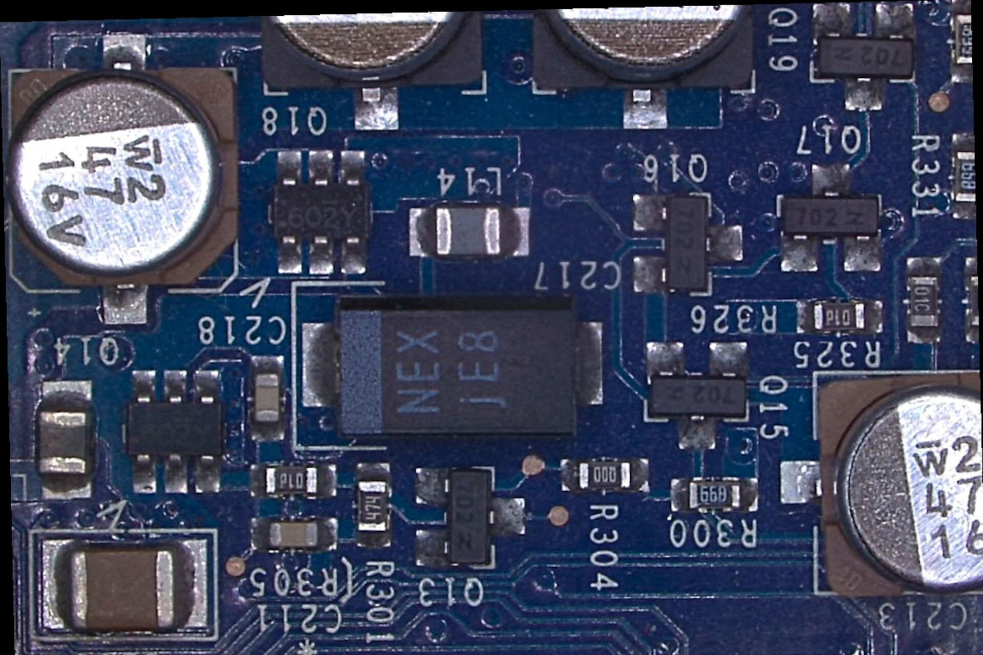
Below is a photo of the DVM6 with a PCBA placed onto its stage, as well as a photo of the PCBA, and a low magnification DVM6 image of a region of it.


Measurement, analysis, and operation with intuitive software
In order to achieve a fast and reliable workflow for QC, FA, and R&D with a digital microscope, an intuitive software package with numerous functions for microscope control, simple yet flexible image acquisition, and sample analysis is essential. For example, the software used with the DVM6, LAS X, offers storage of multiple user profiles for cases where many different users work with the same microscope. The Z-stack function of LAS X enables users to record images in various focal planes over a defined Z-range for a sample feature or the entire sample itself. The extended depth of field (EDOF) mode provides a multi-focus image without the need to set begin nor end levels. Both Z-stack and EDOF allow the fast creation and analysis of 3D topographic surfaces [4] of a sample.
In addition, with LAS X software, users can choose different modes for a large XY scan, such as "mark & find", "tile scan", and "spiral scan". An interactive mode called "Live Image Builder" for XY (2D image), Z (3D image), and XYZ (3D image over extended area) can also be used.
Examples of 3D sample analysis using LAS X are shown below with a PCBA and SMD (surface mounted device) hybrid sample. Report generation with just one click is also explained.
 chips and a solder joint; captured with a DVM6 using the EDOF function of LAS X. See also same image below with color Z-scale.")


.")


Fast magnification change over a large range
The objective lenses of the DVM6 are quickly and easily changed during operation of the instrument with essentially no increase in workflow. A video shows how simple it is to switch objectives [5].
There is a selection of 3 plan apochromatic (chromatic correction over the red, green, and blue wavelengths and flatness correction over the whole field of view) objectives (low, middle, and high magnification). There is also the 16:1 integrated zoom optics with which users can achieve total magnification values from 12x to 2,350x (with the recommended 27 inch (69 cm) diagonal monitor display in accordance with the standard ISO/DIN 18221) [6]. The zoom optics works with each of the 3 objectives for low, middle, and high magnification, making it possible to change the magnification in a continuous manner over the full range.
It must be kept in mind that the final magnification value for digital microscopy will depend on the monitor size used for image display [6]. As mentioned above, it is recommended that the DVM6 be operated with a 27 inch (69 cm) diagonal monitor. Images of an electronic sensor acquired with the DVM6 using the low, middle, and high objective at low, middle, and high magnification are shown below.
.")
.")
.")
Encoded parameters
An instrument with hardware in direct communication with computer software to allow the automatic tracking and saving of specific parameters is called an "encoded" device. Encoding is very useful for rapidly recalling parameters and settings stored during data acquisition. Encoding is invaluable for reproducibility and reliability and also contributes to a more efficient workflow.
For the DVM6, the objective and zoom optics, camera pixel resolution, sample-stage position and rotation angle (whether manual or motorized movement), microscope-head tilt angle, and illumination system settings are encoded and stored via LAS X software. An example of the encoding of some of these parameters during operation of the DVM6 is shown below.
 from objective and zoom optics, microscope-head tilting angle (5°), and stage rotation angle (20°) are encoded.")
 or object field is also encoded (objective and zoom optics).")
Conclusions
Efficient, reliable quality control and assurance (QA/QC), failure analysis (FA), and research and development (R&D) for electronics parts, such as printed circuit boards (PCBs) and assemblies (PCBAs), can be achieved with the DVM6 digital microscope. Here 3 of its benefits were discussed: intuitive software with many functions for microscope operation, image capture, and data analysis; convenient ways to change magnification rapidly over the full range from 12x up to 2,350x; and encoding of all important parameters and settings (optics, camera, stage, head, and illumination) for easy, fast recall. These features allow DVM6 users to acquire and analyze data fast and reliably for QC, FA, and R&D workflow efficiency.
References
- J. DeRose, G. Schlaffer, What You Always Wanted to Know About Digital Microscopy, but Never Got Around to Asking, Science Lab (2015) Leica Microsystems.
- J. DeRose, Digital Inspection Microscope for Industrial Applications: How to choose the right microscope which helps users achieve efficient workflows, Science Lab (2023) Leica Microsystems
- M. Doppler, How to Use a Digital Microscope to Streamline Inspection Processes: Webinar on-demand, Science Lab (2021) Leica Microsystems.
- C. Smith, M. Horz, How To Create EDOF (Extended Depth of Focus) Images: Overview of the 3 methods in the LAS X software, Science Lab (2019) Leica Microsystems.
- Objective change: plug and see, Video DVM6 product page, Leica Microsystems.
- DeRose J.A., Doppler M., What Does 30,000:1 Magnification Really Mean? Some Useful Guidelines for Understanding Magnification in Today’s New Digital Microscope Era. Science Lab (2018) Leica Microsystems.

. With DIC users are able to visualize small height differences on the wafer surface more easily.")

