Contact Us

Julia Roberti , Dr

Julia Roberti

Julia studied Chemistry at the University of Buenos Aires, where she worked on Photochemistry and Analytical Chemistry. She focused on the characterization of luminescence of lanthanide complexes, and on the development of boron quantification methods for boron neutron capture therapy (BNCT) of cancer. After graduating, she moved to Göttingen to carry out her doctoral research at the Max Planck Institute for Biological Chemistry. She developed in vitro and in situ fluorescence labeling strategies to elucidate the oligomerization and aggregation mechanisms of the Parkinson's disease-associated protein alpha-synuclein. In 2012, she joined EMBL as Humboldt postdoctoral fellow, and applied advanced confocal microscopy and nanoscopy imaging to study chromatin compaction at the interphase-to-mitosis transition. She joined Leica Microsystems in 2017 as Product Manager for advanced confocal imaging.
Insights into Vesicle Trafficking

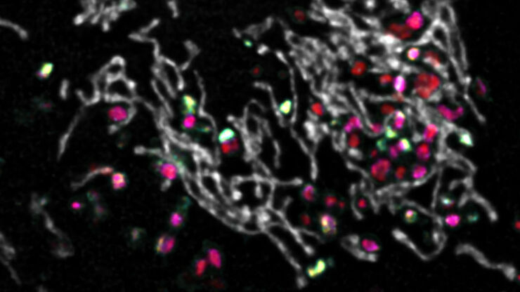
STELLARIS provides integral access to complementary layers of information for dynamic, structural, and mechanistic insights into vesicle trafficking.
Spectral separation of 11 fluorophores coupled to polystyrene beads on a STELLARIS confocal system.

Multiplexing through Spectral Separation of 11 Colors

Fluorescence microscopy is a fundamental tool for life science research that has evolved and matured together with the development of multicolor labeling strategies in cells tissues and model…

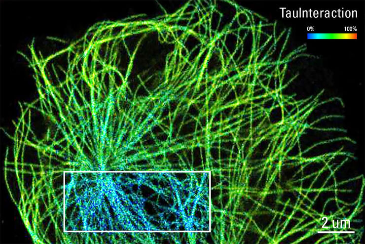
TauInteraction – Studying Molecular Interactions with TauSense

Fluorescence microscopy constitutes one of the pillars in life sciences and is a tool commonly used to unveil cellular structure and function. A key advantage of fluorescence microscopy resides in the…

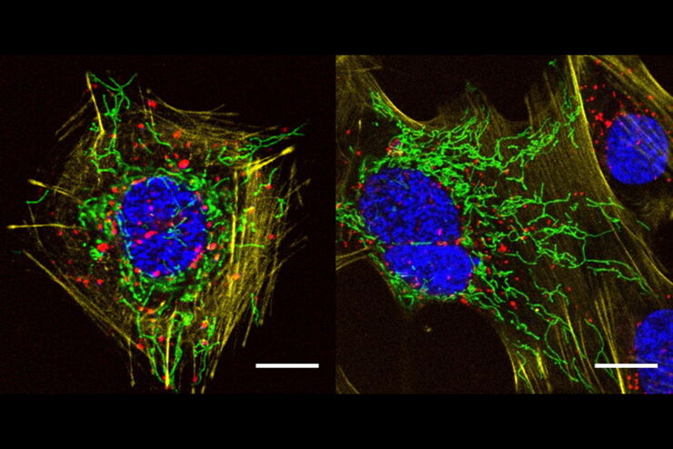
A Versatile Palette of Fluorescent Probes

Researchers at the Max Planck Institute for Medical Research in Heidelberg have developed a general strategy to synthesize live-cell compatible fluorogenic probes, and the result are the new MaP (Max…

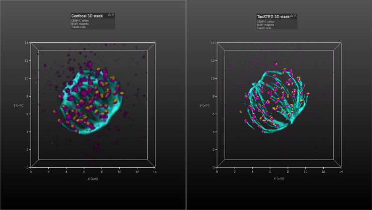
Kinetochore Assembly during Mitosis with TauSTED on 3D

Three-dimensional organization of the mitotic spindle together with the distribution of CENP-C and BUB1 based on TauSTED with multiple STED lines (592, 660 and 775 nm) can provide insights on…

Observing Malaria Infection at the Right Spot in the Human Host

Malaria is a life-threatening disease transmitted through the bites of mosquitoes infected with protozoan parasites. The most common and dangerous type of malaria is caused by the parasite Plasmodium…
Scroll to top