
Cataract surgeons rely on a red reflex
The red reflex is the reddish-orange reflection from the retina that is observed through an ophthalmic microscope as coaxial light goes into and out of the pupil of the eye. Cataract surgeons rely on the red reflex and depth of field to precisely view the elements in the eye during each step of surgery, such as corneal incisions, division of nuclear material, removing the cortex, cleaning of the anterior and posterior leaves of the capsular bag, placing the IOL.
Consistent visualization is especially important when performing capsulorhexis which is best judged by the shadow its edge creates with the red reflex. The brighter and more even the red reflex is, the more distinct the capsular edge becomes. This enhances surgical ease in performing centered and appropriately sized capsulorhexis in all types of cataract.
Consequences of a diminished red reflex
As the eye moves or the position of the microscope is changed, the red reflex may decrease, making certain phases of cataract surgery more difficult, even dangerous. For example, with a diminished red reflex it is more difficult to create a round and appropriately sized capsulorhexis, and to determine where within the cataract the phaco tip is being placed.
Most notably, without consistent illumination it is more difficult to view remaining nuclear material in the capsular bag during phacoemulsificaton and the posterior capsule bag during the procedure.
Four beam paths facilitate stable red reflex
A new technology for an ophthalmic microscope makes it possible to work with a stable and consistent red reflex. Four individual coaxial illumination beam paths down to the patient eye with a perpendicular entrance to the retina are providing a straight red reflex throughout the entire procedure (Fig. 1).
Compared to known microscopes, the image appears equal for all possible observers, the main observer, the assistant and the documentation.

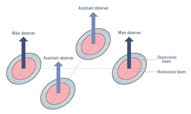
Stereo red reflex for all observers
The CoAx4 Illumination makes the red reflex fully visible for both main surgeon and assistant. Surgeon and assistant sharing the same view of the surgical field with excellent contrast and red reflex makes cataract surgery more secure and is a valuable asset to teaching (Fig. 2).
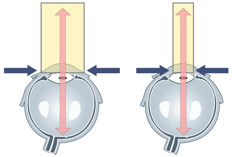
Adjustable illumination diameter enhances contrast
The Proveo 8 ophthalmic microscope is the first system to feature the new technology. With an adjustable illumination diameter sizeable from 4 – 23 mm the surgeon can choose via footswitch if he needs to illuminate the whole eye or prefers to match the field of illumination to the pupil size. A reduced illumination diameter produces less sclera reflections and offers more contrast, while a wider illumination is more flexible with respect to patient’s movements (Fig.3).
The CoAx4 Illumination also provides a camera image that is identical to the optical view through the ocular. The light for the camera is taken from the assistant optical path and thus leaves the main surgeon’s view with 100% of the light.