
Introduction
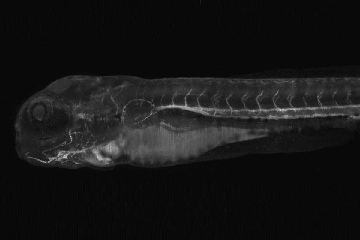
In light sheet microscopy, specimen preparation is a crucial topic. Ideally, it should be as simple and fast as possible. The DLS system has the advantage that it allows for horizontal mounting and easy sample acces. Specimen preparation has only two requirements: the sample needs to be placed in the focal plane of the detection objective (between the TwinFlect mirrors) and slightly elevated from the petri dish bottom, so that the mirrors/light sheet can reach the distal part of the sample.
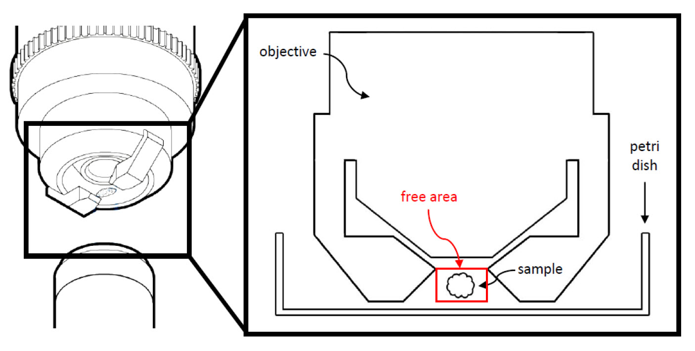
U-shaped glass capillaries, which are customized for different TwinFlect mirror sizes, help users fulfill these specimen preparation requirements in an intuitive manner.

Workflow
Materials and methods
U-shaped capillaries are available in two different sizes:
- Outer diameter = 1.5mm; inner diameter = 1.03mm, length 20mm (Order number 158007061, 50 capillaries) → fit TwinFlect 2.5mm, TwinFlect 5mm and TwinFlect 7.8mm
- Outer diameter = 2.5mm; inner diameter = 2.04mm, length 20mm (Order number 158007060, 50 capillaries) → fit TwinFlect 5mm and TwinFlect 7.8mm
With a length of 20mm, the u-shaped glass capillaries are ideal for use with 35mm glass bottom petri dishes (e.g. Greiner, ibidi, Matek,…).

-b-poly(isoprene). Right: Poly(styrene)-b-poly(methyl methacrylate).")

