Contact Us

Philipps University Marburg, Institute of Cytobiology and Cytopathology, Germany

Philipps University Marburg, Institute of Cytobiology and Cytopathology, Germany

The Institute is integrated into the Faculty of Medicine of the Plilipps University in Marburg. The members are doing basic research in cell biology and biological chemistry. The institute is also responsible for the training of students of medicine, dentistry and human biology. Central research projects deal with molecular mechanisms in the biogenesis of cell organelles. The understanding of mutations of the involved proteins is of particular interest here, because these cause diseases. The research is mainly done using the model organisms yeast, mouse and rat. Further, different cell culture systems from mammals are used. The spectrum of methods in the particular projects involves cell biological, biochemical, immunohistochemical, molecular biological and genetical techniques. 

Central research areas are:

  • Biogenesis of Ferric-Sulfur-Proteins in the Mitochondria and in the Cytosol
  • Molecular Basics of the neurodegenerative Disease Friedreich's Ataxie
  • Mechanism and Regulation of the Mitochondrial Ferric-Transport
  • Maturation and Sorting of Proteins in Polarized Epithelia Cells
  • Function of lysosomale Proteins
  • Function of the Mys-Binding Protein Miz-1 in Keratinocytes
http://www.uni-marburg.de/fb20/cyto/

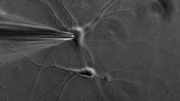
Patch pipette touching a murine hippocampal neuron. Image courtesy of A. Aguado, Ruhr University Bochum, Germany.

What is the Patch-Clamp Technique?

This article gives an introduction to the patch-clamp technique and how it is used to study the physiology of ion channels for neuroscience and other life-science fields.
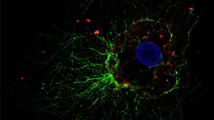
Cellular microtubule network in a fibroblast cell

How to Prepare your Specimen for Immunofluorescence Microscopy

Immunofluorescence (IF) is a powerful method for visualizing intracellular processes, conditions and structures. IF preparations can be analyzed by various microscopy techniques (e.g. CLSM,…
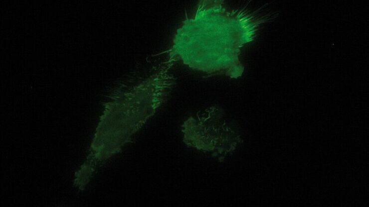
TIRF image of brest carcinoma tumor cells expressing GFP tagged cell adhesion Molecule CD44 that is expressed on the cell membrane, imagined in TIRF.

Total Internal Reflection Fluorescence (TIRF) Microscopy

Total internal reflection fluorescence (TIRF) is a special technique in fluorescence microscopy developed by Daniel Axelrod at the University of Michigan, Ann Arbor in the early 1980s. TIRF microscopy…
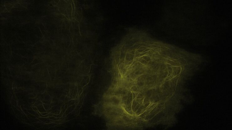
TIRF Image of Tubulin, YFP, penetration depth: 120 mm

Applications of TIRF Microscopy in Life Science Research

The special feature of TIRF microscopy is the employment of an evanescent field for fluorophore excitation. Unlike standard widefield fluorescence illumination procedures with arc lamps, LEDs or…

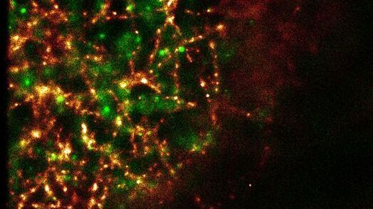
Super-Resolution GSDIM Microscopy

The nanoscopic technique GSDIM (ground state depletion microscopy followed by individual molecule return) provides a detailed image of the spatial arrangement of proteins and other biomolecules within…
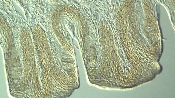
Section taste buds rabbit, differential interference contrast microscope

Optical Contrast Methods

Optical contrast methods give the potential to easily examine living and colorless specimens. Different microscopic techniques aim to change phase shifts caused by the interaction of light with the…
Tartaric acids, polarization

Polarization Contrast

Polarization microscopy is routinely applied in material sciences and geology to identify minerals on the basis of characteristic refraction properties and colors. In biology, polarization microscopy…
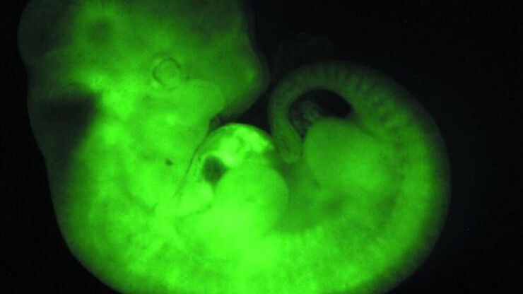
Transgenic Mouse Embryo, GFP

Fluorescence in Microscopy

Fluorescence microscopy is a special form of light microscopy. It uses the ability of fluorochromes to emit light after being excited with light of a certain wavelength. Proteins of interest can be…
Scroll to top