Logo Leica Microsystems
  • Products
    Light Microscopes
    InvertedUprightPhase ContrastDarkfield
    Stereo Microscopes & Macroscopes
    MacroscopesRoutine ManualResearchDissectionMaterial AnalysisInspectionDIC
    Automated Microscopes
    Confocal Microscopes
    Microhub - Mica
    THUNDER Imaging Systems
    Surgical Microscopes
    Digital Microscopes
    Used Microscopes & Imaging Equipment
    Sample Preparation for Electron Microscopy
    Sputter Coater & Freeze FracturingIon Beam MillingUltramicrotomes & Cryo-Ultramicrotomes
    Microscope Cameras
    Microscope Software
    Microscope Objective Lens
    ObjectivefinderObjective ClassesLabeling of ObjectivesLeitz Optic Center
    Microscope Parts & Accessories
    CamerasIlluminationErgonomicsIncubation Systems and StagesExternal Filter WheelsEyepieces and Graticules
    Product Archive
  • Solutions
    Life Science
    Cryo-Electron TomographyCancer ResearchCell BiologyCell CultureFluorescenceFluorescence Lifetime ImagingLaser MicrodissectionLive Cell ImagingMicromanipulationModel Organisms in ResearchNeuroscienceOrganoids and 3D Cell CulturePhotomanipulationSuper-ResolutionVirologyZebrafish Research
    Industry & Manufacturing
    Technical CleanlinessAutomotive & TransportationEnergy, Mining and Natural ResourcesJewelry Making / Hand EngravingMetals & Machine EngineeringElectronics & Semiconductor IndustryMedical Device Quality Assurance & ControlSteel Quality RatingMetallographyWatchmaking Industry
    Medical
    Cataract SurgeryOptical Coherence Tomography (OCT)NeurosurgeryOphthalmologyENTPlastic & Reconstructive SurgeryDental Microscopy
    Education
    Earth Science EducationForensic Science EducationLife Science EducationMaterials Science EducationWi-Fi Education
    Materials & Earth Science
    Sample Preparation for Materials ScienceArt Conservation
    Microscopy in Pathology
    Clinical PathologyAnatomic Pathology
    Forensic Science
    Pharmaceutical Engineering Research
  • Service
  • Science Lab
  • Company
    About us
    About DanaherIntegrity and ComplianceWhat we stand forLeadership CornerOur HistoryIdeation
    News
    Events
    MicaCam Livestream
    Careers
    Certificates
    Product Security
    Product Security Updates
    Quality Policy
    Terms and Conditions
  • EN
    DE FR ES IT PT 中文 日本語 한국어
  • Contact
  • Contact
  • Contact us online
  1. Home
  2. Science Lab
  3. Institutions

University of Utrecht, The Netherlands

www.uu.nl

Tags

  • Application Note
  • EM Sample Preparation
  • Sample Preparation
  • Electron Microscope
  • SEM
  • STED image of peripheral section of HeLa cell nucleus
    STED

    Abstracts of the 7th European Super-Resolution User-Club Meeting

    The 7th Super-Resolution User Club Meeting was held in collaboration with Prof Pavel Hozák , at the Institute of Molecular Genetics of the ASCR in Prague. Keeping the event close to science is one of…
    Read article
    Dec 17, 2017
    Story
  • Live Cell Imaging

    Five Questions Asked: Prof. Dr. Jacco van Rheenen speaks about the most important considerations when imaging deep into mouse tissue

    When operating a confocal microscope, or when discussing features and parameters of such a device, we inescapably mention the pinhole and its diameter. This short introductory document is meant to…
    Read article
    May 19, 2017
    Story
  • EM Sample Preparation

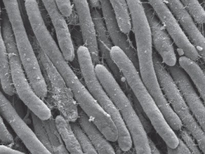
    Bacteria Protocol - Critical Point Drying of E. coli for SEM

    Application Note for Leica EM CPD300 - Critical point drying of E. coli with subsequent platinum / palladium coating and SEM analysis. Sample was inserted into a filter disc (Pore size: 16 - 40 μm)…
    Read article
    Oct 13, 2016
    Story
  • Application Note

    Structural Study of C. elegans

    Application Note for Leica EM ICE, Leica EM AFS2 - Wildtype L4 stage C. elegans (N2 strain) were placed in the 100 μm deep side of Lecithin-coated (see detailed protocol*) type A 3 mm Cu/Au carriers…
    Read article
    Sep 16, 2016
    Story
  • Application Note

    Cultured Rat Hippocampal Neurons

    Application Note for Leica EM ICE - Rat Hippocampal neurons, cultured on 50 μm thick Aclar (Aclar embedding film, EMS) for 19 days, were frozen in the 100 μm deep side of lecithin coated (detailed…
    Read article
    Aug 29, 2016
    Story
  • EM Sample Preparation

    Black Mould Protocol

    Application Note for Leica EM CPD300 - Critical point drying of Black mould (Aspergilus niger) with subsequent platinum / palladium coating and SEM analysis to detect conidiospores. Sample was…
    Read article
    Aug 09, 2016
    Story
  • Electron Microscope

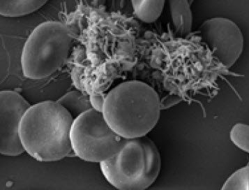
    Human Blood Cells Protocol

    Application Note for Leica EM CPD300 - Life Science Research. Species: Human (Homo sapiens) Critical point drying of human blood with subsequent platinum / palladium coating and SEM analysis.
    Read article
    May 24, 2016
    Story
Logo Leica Microsystems
  • © 2022 Leica Microsystems
  • Terms and Conditions
  • Imprint
  • Terms of Use
  • Privacy Policy
  • Do not sell my personal data
  • Product Security
  • Cookie Settings
  • Language:English
    DeutschEnglishEspañolFrançaisItalianoPortuguês日本語한국어中文