Introduction
Diseases which can cause fibrosis, e.g. severe asthma, ischemic heart disease, cirrhosis of the liver, end stage kidney disease, and idiopathic pulmonary fibrosis (IPF), involve the inappropriate formation of scar tissue in an internal organ [1]. Injuries to the tissue, such as particulate matter or toxins in the lungs, initiate an inappropriate and unnecessary wound healing response leading to organ failure and death. The mechanism behind fibrosis is poorly understood.
Many secreted and cell-surface mammalian proteins are glycosylated and many of the glycosylation structures have sialic acids as the monosaccharide at the distal tip or tips of the polysaccharide [1]. Some viruses, bacteria, protozoa, and all mammals have sialidases (also known as neuraminidases) that remove the sialic acids from glycoconjugates.
To better understand the mechanism behind fibrosis that drives the formation of increasing amounts of scar tissue, fibrotic lesions in human and mouse lung tissue were studied with the goal of finding relationships between extensive desialylation of glycoconjugates and upregulation of sialidases. The sialidase inhibitors DANA and oseltamivir (Tamiflu) were used to study conditions related to pulmonary fibrosis in the mouse bleomycin model [1].
The non-fibrotic and fibrotic regions of collagen in mouse lung tissue were imaged with brightfield and polarized light microscopy in this study to determine if they could be distinguished more easily.
Challenges when imaging fibrotic regions of collagen
For the study of collagen in lung tissue and pulmonary fibrosis, it is difficult to distinguish between fibrotic and non-fibrotic regions of the tissue with staining and brightfield microscopy only.
Methods
The DM6 B upright microscope with brightfield and polarized illumination was used for image acquisition. The mouse lung section specimen was stained with Sirus red for collagen in both fibrotic and non-fibrotic regions. A motorized stage and the LAS X Navigator software allowed a much larger area of the specimen to be imaged via mosaic imaging and image stitching [2]. Many high-resolution images were acquired of the area of interest, enabling much easier statistical analysis.
Results
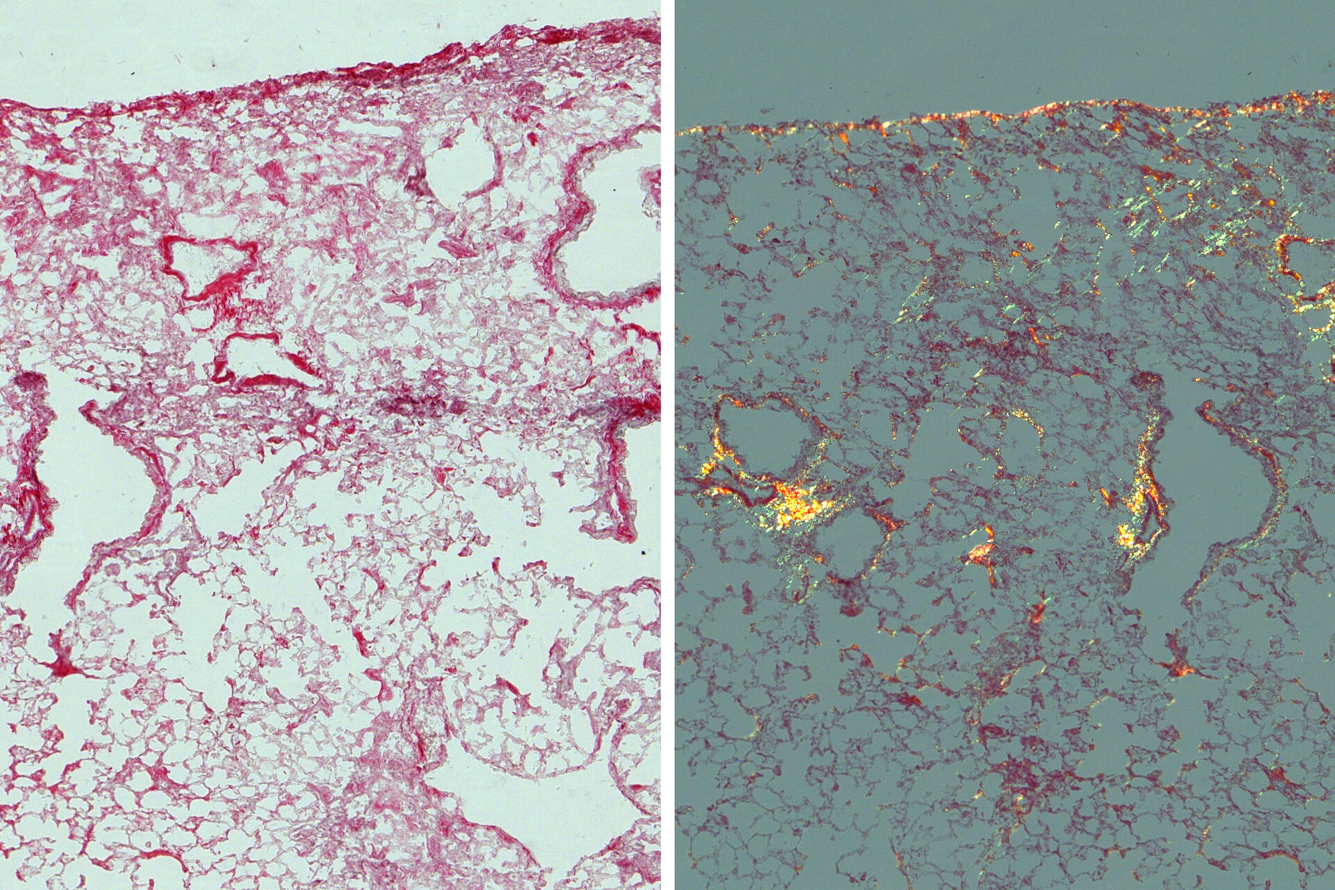
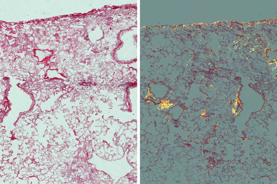
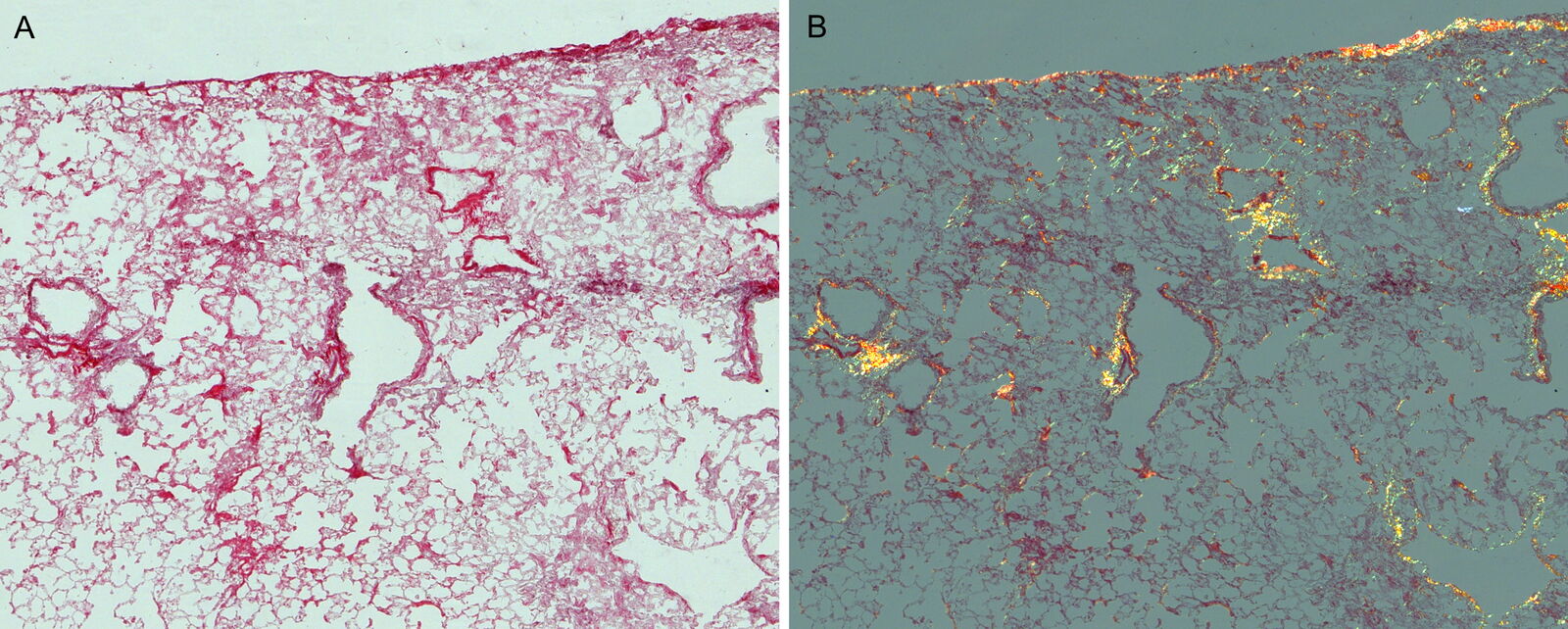
Images acquired of the mouse lung sections are seen in figure 1. With regular brightfield imaging (Fig. 1A), all collagen appeared as red and could not be distinguished between fibrotic and non-fibrotic regions of the lung specimen. With polarized light (Fig. 1B), there is a notable difference in color (red versus yellow) for the collagen between the 2 regions which cannot be seen with brightfield.


 which was imaged with extended depth of field (EDOF) using digital microscopy.")