A reliable diagnosis is the basis of a successful treatment
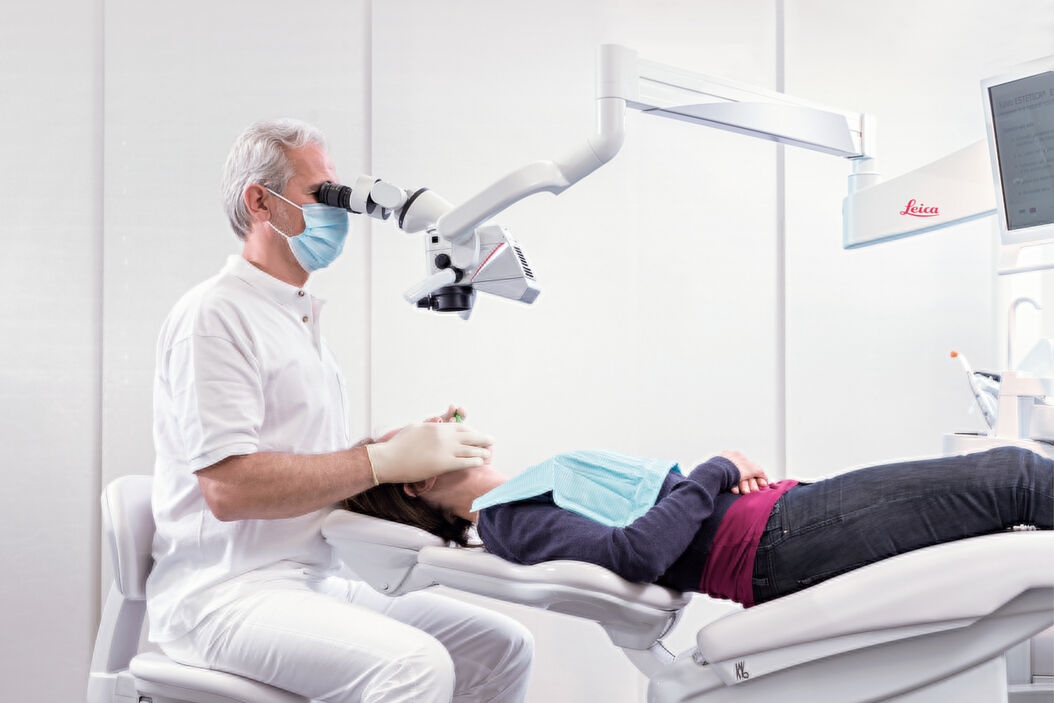
A dental microscope such as the Leica M320 can provide significant visualization benefits at all stages of endodontic treatment, from diagnosis and exposure of the access cavity to preparation, and from three-dimensional obturation to postendodontic management. High magnification, large depth of field and bright, coaxial illumination of the surgical field can reveal fine details, helping the dentist to manage difficult cases with increased precision.
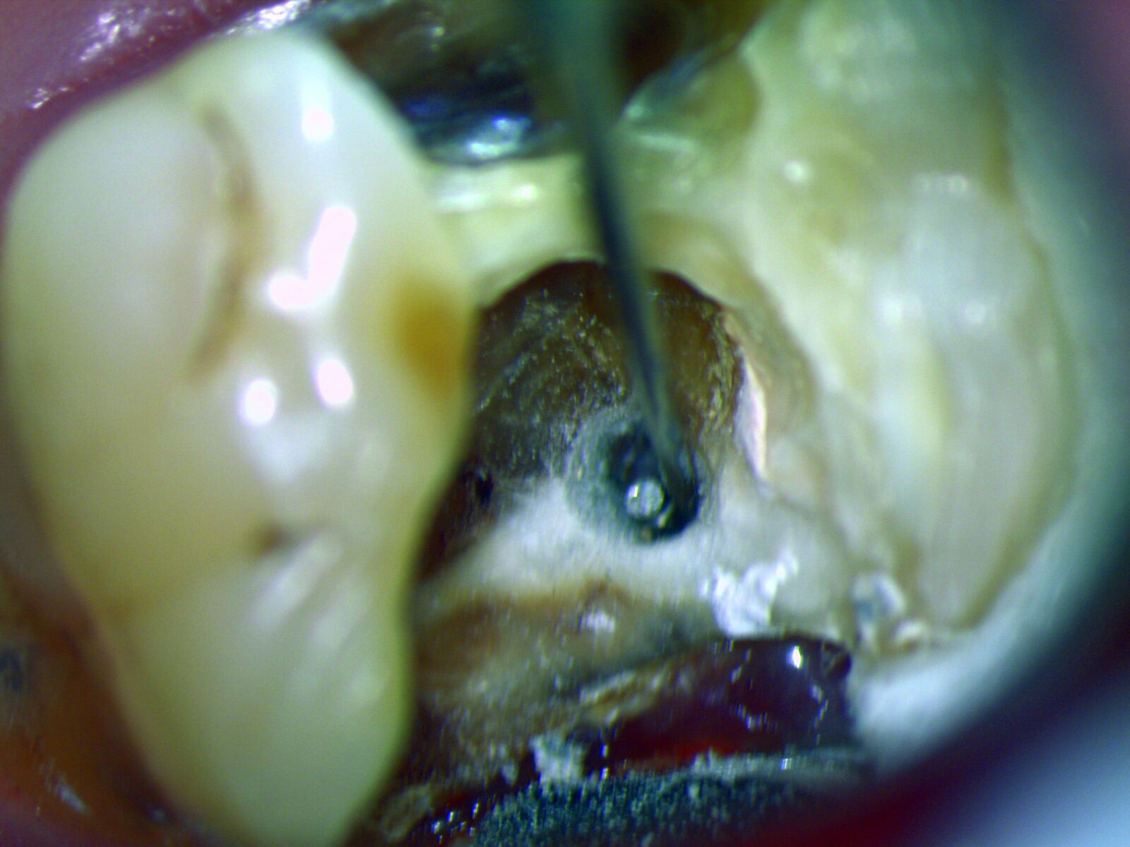
Successful therapy starts with an accurate diagnosis. The dental microscope can reveal microfractures or longitudinal fractures that are easily overlooked, but are often a cause for pain and demand specific treatment planning. One of the main reasons for endodontic failure is untreated root canal structures with remaining bacterial infection. The dentist therefore needs to be able to identify the whole root canal structure before treatment. Due to its high magnification and coaxial LED illumination, a dental microscope such as the Leica M320 enables visualization of the root canal all the way down to the apex. Anatomical variations like three-rooted premolars or c-shaped canals are thus clearly visible and can be adequately treated.
Better visualization of obliteration and denticles
A considerable number of patients show signs of obliterations, due to the rising age of population, or calcifications, including denticles. These cannot be seen with the naked eye, but may block the root canal entrance and impair instrumentation. The dental microscope helps to visualize these obstacles so they can be removed or dissolved preparing a suitable treatment for these patients.
The microscope lowers the risk of instrument breakage."
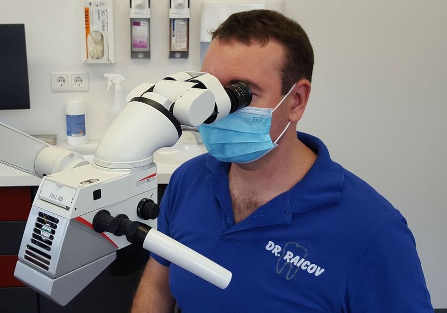
Root canal treatments require the use of the finest surgical instruments. Such delicate instruments can potentially fracture inside the narrow root canal leaving behind fragments which can cause new inflammations and severe pain, calling into question the attempt to save the tooth. The complete removal of these fragments depends primarily on sharp visualization; if the fragment can be removed without any further loss of tooth structure, the prognosis for preservation of the tooth is quite good. In addition, improved visualization may help avoid instrument breakage in the first place. “I also think that the microscope lowers the risk of instrument breakage as you can create much better root canal access and observe the instruments’ movement more reliably”, says Dr. Dean Raicov, endodontist in Germany who works with a Leica M320 dental microscope.
Read the full interview with Dr. Raicov: “The Dental Microscope in Endodontics”.
Improved prognosis for special therapies at the apex
Modern therapies concerning the apex – like sealing an open apex (apexification) or removing an infected apex (apicoectomy) – demand special treatment techniques that are aided by the use of a dental microscope. Thanks to high magnification along with the use of microsurgical equipment, the size of the access cavity through the bone to the root tip can be minimized. This results in a less traumatic procedure, with less pain and potentially fewer complications. Microscope supported therapies also result in improved safety as surrounding structures, such as nerves and roots of neighboring teeth, are less likely to be affected.
Facilitating microsurgical techniques
With the development of new microsurgical instruments and the full-time use of a dental microscope, endodontic surgery has entered into a microsurgical era. The surgeon can now visualize the entire bony crypt, lesion, and root tip securely and reliably. Microsurgical operating techniques tend to be more successful and less traumatic for patients. On the one hand, increased precision helps to preserve those teeth that would otherwise be lost. On the other hand, the dental microscope clearly reveals major obstacles for a successful restoration of the tooth, such as internal cracks extending down the canals, massive perforation, or inadequate coronal structure. The patient thus benefits from avoiding unneeded treatment, including pain and costs, if the tooth is not salvageable.

The benefits of the Leica M320 dental microscope at a glance
- Premium apochromatic optics, high magnification and two LEDs deliver clear, bright, true-to-life color visualization
- Five-step magnification changer helps minimize workflow interruption
- The optional multifocal objective allows working distance to be quickly and easily adjusted between 200 mm and 300 mm during examination and treatment.
- Flexible, ergonomic design and customized ergonomic accessories for adaptation to body frame and clinic set up.
- Optional full HD camera to demonstrate procedures via video or live image, train the team or share experiences with peers.
Related Articles
-

Dental Loupes vs Microscopes: Exploring Visualization Options in Dentistry
Dental professionals often ask: “Should I use dental loupes or invest in a microscope?” This article…
Jun 22, 2026Read article -

Six Features to Consider when Choosing a Dental Microscope
The dental surgical microscope has become increasingly important for high-quality and successful…
Jun 02, 2026Read article -

Improving Ergonomics With a Dental Microscope
Here, we show you how Dr. David Blanc, a dental surgeon and ergonomics consultant, improves physical…
Apr 07, 2022Read article
Related Pages
-
Surgical Microscopes
The surgical microscopes of Leica Microsystems are exactly geared to the requirements of…
Visit related page
