Fluorescence lifetime as a probe for the nano-environment
This lifetime is a characteristic parameter of each fluorescent dye or molecule that may change with its nanoscopic surroundings or its conformational state. Lifetime information probes the molecular environment for its composition, such as ion concentration, pH, lipophilicity, or the binding to other molecules.
Combination of lifetime measurement and imaging
FLIM combines lifetime measurements with imaging: lifetimes obtained at the pixel-level are color-coded to produce images. Thus, FLIM delivers information about the spatial distribution of a fluorescent molecule together with information about its nano-environment. This way an additional dimension of information is obtained.
Principle of FLIM data acquisition and analysis
- Repeated measurement of the time between the laser excitation of the fluorophore and fluorescence photon arrival at each pixel of a detector (refer to figure 1A).
- Calculation of a histogram of photon counts versus arrival time after the laser pulse.
- Fit of the exponential decay curve, F(t) = A0 exp (-t/τ), to each histogram. The amplitude, F(t), reflects the total number of photons detected at a specific time and the time constant, τ, is called the fluorescence lifetime (refer to figure 1B).
- Lifetime images are displayed using an arbitrary color for each lifetime value (refer to figure 1C).
 Diagram showing the repeated measurement of the fluorescence photon arrival time")
Solution for FLIM
Harness the power of FLIM with the STELLARIS 8 FALCON system. Efficiently investigate cellular physiology and explore dynamics in living cells.
![Intensity and FLIM image of U2OS cells immunostained with α-tubulin (AF546) and vimentin (AF555) [1]. The scale bar represents 5 µm and the color bar 0–3 ns.](/fileadmin/_processed_/1/9/csm_what-is-flim-fig-2_251c8d2ce7.jpg "Intensity and FLIM image of U2OS cells immunostained with α-tubulin (AF546) and vimentin (AF555) [1]. The scale bar represents 5 µm and the color bar 0–3 ns.")
 and acceptor (A) molecule which participate in FRET (Förster resonance energy transfer).")

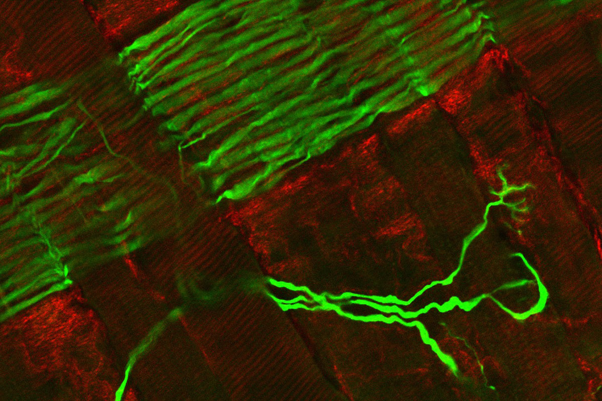
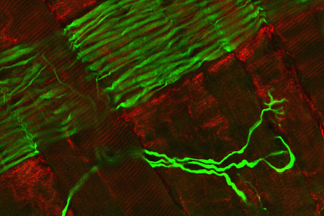
, Tropomyosin (cardiomyocytes, red) and GFP (primordial cardiac layer, green).")