Cell Biology Research
When your research is centered on understanding the cellular basis of human health and disease, it is critical to investigate the cells of interest in spatiotemporal and molecular detail. Consequently, microscopy is an ever-important tool in cell biology, allowing you to study your specimens in detail within their structural environment, as well as, analyze cellular organelles and macromolecules. Cell biology imaging is done with a range of light and electron microscopes. Imaging solutions offered by Leica Microsystems are designed to maximize your cell biology research.
Contact a local specialist for advice on the right cell biology system for your needs.
Challenges with cellular imaging
The challenges faced when using microscopy for cell biology research vary, as the imaging of inter- and intra-cellular events requires a range of samples that vary in size and complexity. Imaging of these events needs to be performed from the nanometer to the millimeter scale.
Additionally, the study of cells under the microscope is further influenced by whether they are live or fixed samples, as these cases pose different imaging challenges. One challenge is how to capture fast dynamic events with the right resolution. Another one, for any cellular structures or events that do not produce a natural contrast during imaging, is choosing the right fluorescent proteins, antibodies, or nucleic acid probes to use as tags for specific proteins, DNAs, and RNAs in the cell to be viewed with fluorescence microscopy.
Finding the right solution for cellular imaging
Choosing the right microscopy method is critical if you want to maximize your research output and obtain the highest quality data.
Leica Microsystems offers various solutions to improve your cell biology research. These range from digital imagers that enhance daily cell culture tasks to high-end imaging solutions that enable the study of single molecules in detail or maximize the amount of results you get from your sample.
THUNDER Imager
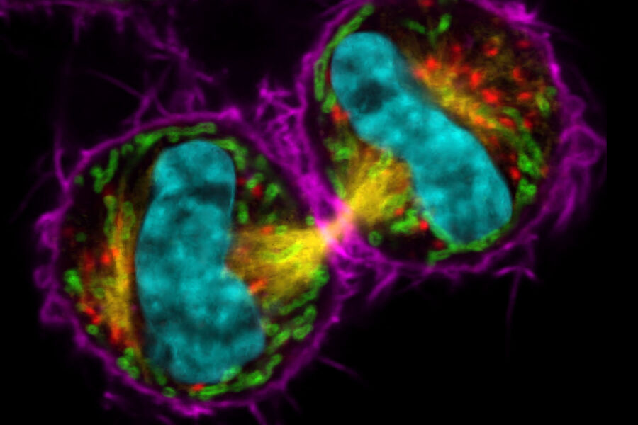
THUNDER Imagers provide you with a solution for advanced 3D cell culture assays, whether you want to study stem cells, spheroids, or organoids. THUNDER Imager 3D Live Cell & 3D Cell Culture meets your requirements to study cells close to their physiological state by optimizing experimental conditions, e.g., the lowest light intensity and shortest exposure times possible.
Infinity TIRF
TIRF is ideal for imaging dynamic processes and the method of choice for visualizing single molecules with super-resolution.
STELLARIS
Experience the power to see more within your cells, with enhanced image quality, the possibility to image more markers within one sample at the same time, and the capacity to make detailed observations without influencing or damaging your sample with STELLARIS confocal microscope platform.
Cell biology related products
Filter by Area of Application

STELLARIS
With the STELLARIS confocal platform, we have re-imagined confocal microscopy to get you closer to the truth.

THUNDER Imager EM Cryo CLEM
The THUNDER Imager EM Cryo CLEM enables precise identification of cellular structures and smooth, secure transfer of coordinates, images, and samples through your correlative workflow.

K5
sCMOS Microscope Camera

ARTOS 3D
Ultramicrotome for array tomography

Infinity TIRF
Multicolor advanced TIRF module
Featured image
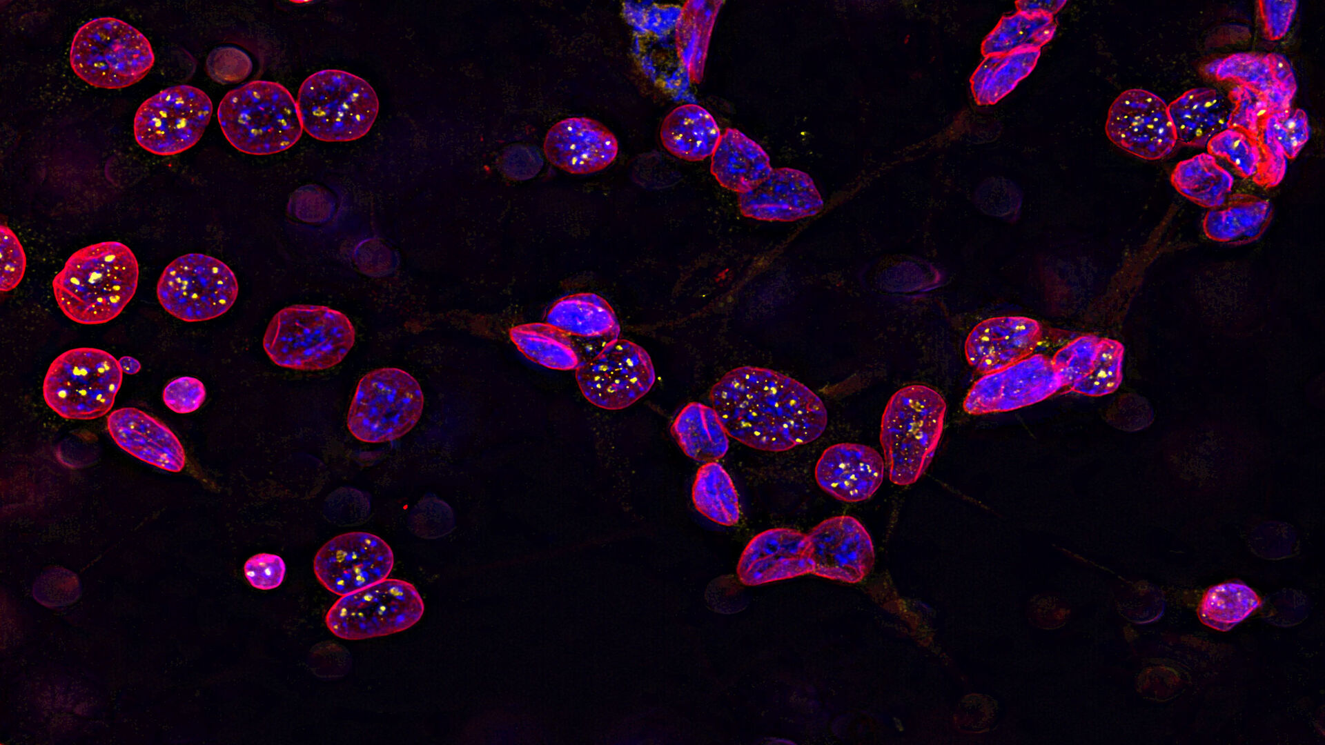
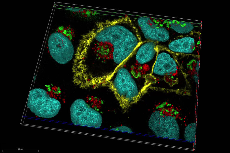
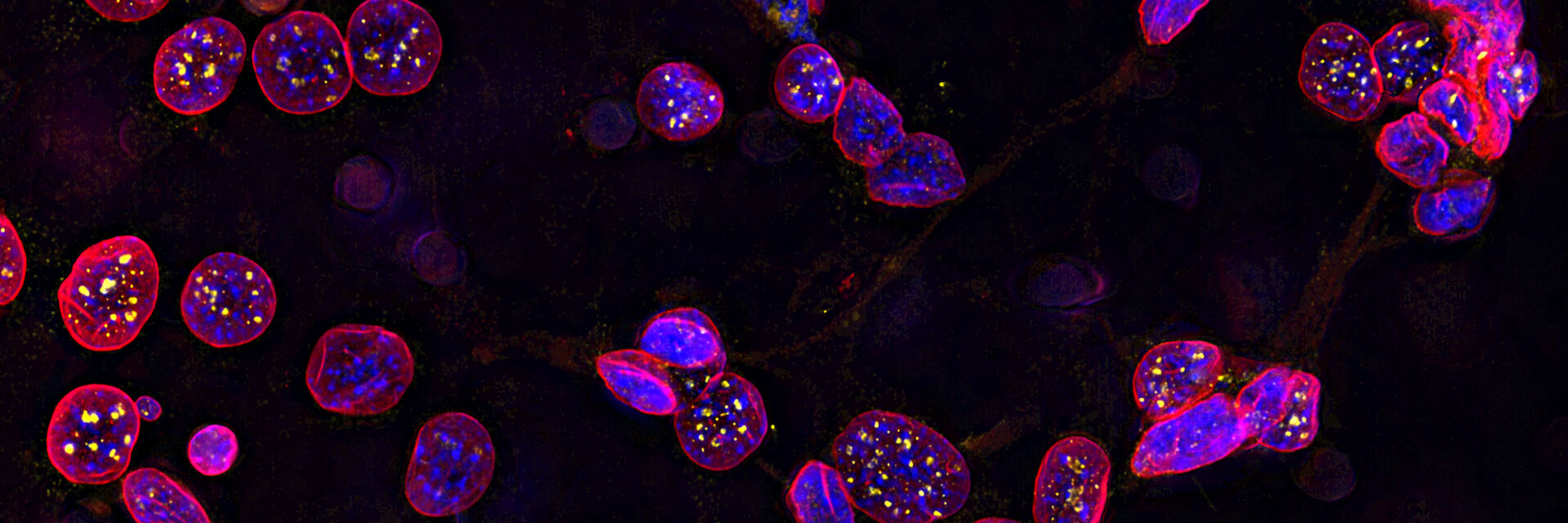
Image of C2C12 cells
The cells are stained with lamin B (magenta) which indicates nuclear structure, Hoechst (blue) indicating DNA, and γH2AX (yellow) indicating damage to DNA. Cells were imaged using a THUNDER Imager 3D Live Cell with a 63X/1.4 oil immersion objective.
Follow us on Instagram
Cell biology related articles
Vibrational Spectroscopic Imaging in Cells, Tissues and Model Organisms
Here we present a webinar on the emerging applications of Stimulated Raman Scattering Microscopy, covering the following topics: Quantitative spectroscopic imaging of label-free or chemically tagged…Read articleNew Light Shed on the Nanodomain Organization of the Endoplasmic Reticulum (ER)
The endoplasmic reticulum (ER) is a continuous membrane organelle in charge of protein synthesis, lipid synthesis and detoxification. The ER structure is described in terms of smooth peripheral…Read articleImaging Intracellular Temperature using Fluorescence Lifetime Imaging Microscopy (FLIM)
This video shows the presentation of Dr. Kohki Okabe given at the 9th Congress of the Federation of Asian and Oceanian Physiological Society (FAOPS) held in Kobe, Japan on 30 March 2019. Dr. Okabe…Read articleDevelopment of fluorescence lifetime imaging microscopy (FLIM) and its relevance for functional imaging
Prof. Ammasi Periasamy, Director, Keck Center for Cellular Imaging, University of Virginia, was interviewed by Dr. Giulia Ossato, Product Manager functional imaging, during Leica Microsystems Meets…Read articleConfluency Check with PAULA Cell Imager
Many cell-based experiments require cells in a certain state. This can include their morphology, fluorescent protein expression, and the confluency of the cell layer. Researchers check their cells…Read articleKaggle Competition for Multi-label Classification of Cell Organelles in Proteome Scale Human Protein Atlas Data
The Cell Atlas, a part of the Human Protein Atlas, was created by the group of Prof. Emma Lundberg at the SciLifeLab, KTH Royal Institute of Technology, in Stockholm, Sweden. Currently, she is a…Read articleImage Gallery: Tunable Multicolor Deep In Vivo Imaging
Today’s life science research focusses on complex biological processes, such as the causes of cancer and other human diseases. A deep look into tissues and living specimens is vital to understanding…Read articleCalifornia NanoSystems Institute at UCLA Publications
A list of the published scientific articles which include work done in the ALMS/MSI Facilities.Read articleIntroduction to Mammalian Cell Culture
Mammalian cell culture is one of the basic pillars of life sciences. Without the ability to grow cells in the lab, the fast progress in disciplines like cell biology, immunology, or cancer research…Read articleFive Questions Asked: Prof. Dr. Jacco van Rheenen speaks about the most important considerations when imaging deep into mouse tissue
When operating a confocal microscope, or when discussing features and parameters of such a device, we inescapably mention the pinhole and its diameter. This short introductory document is meant to…Read articleHuman NK Cell Development Requires CD56-mediated Motility and Formation of the Developmental Synapse
While distinct stages of natural killer (NK) cell development have been defined, the molecular interactions that shape human NK cell maturation are poorly understood. Here we define intercellular…Read articleActin-Dependent Vacuolar Occupancy of the Cell Determines Auxin-Induced Growth Repression
The cytoskeleton is an early attribute of cellular life, and its main components are composed of conserved proteins. The actin cytoskeleton has a direct impact on the control of cell size in animal…Read articleVariety of RNAs in Peripheral Blood Cells, Plasma, and Plasma Fractions
Human peripheral blood contains RNA in cells and in extracellular membrane vesicles, microvesicles and exosomes, as well as in cell-free ribonucleoproteins. Circulating mRNAs and noncoding RNAs, being…Read articleThe Bif-1-Dynamin 2 Membrane Fission Machinery Regulates Atg9-Containing Vesicle Generation at the Rab11-Positive Reservoirs
Application example of HyVolution Super-Resolution - Atg9 is a multispanning transmembrane protein that is required for autophagosome formation. During autophagy, vesicles containing Atg9 are…Read articleSuper-Resolution Optical Microscopy of Lipid Plasma Membrane Dynamics
Plasma membrane dynamics are an important ruler of cellular activity, particularly through the interaction and diffusion dynamics of membrane-embedded proteins and lipids.