Life Science Research

Life Science Research

Life Science Research

Leica Microsystems’ life science research microscopes support the imaging needs of the scientific community with advanced innovation and technical expertise for the visualization, measurement and analysis of microstructures.
Brain organoids labeled with lamin (green) and tubulin (magenta), acquired using Viventis Deep Dual View Light Sheet Microscope. Courtesy of Akanksha Jain, Treutlein Lab ETH-DBSSE Basel (Switzerland).

Advanced Imaging Solutions for Biopharma

Advanced imaging solutions empower biopharma researchers to uncover critical insights in drug discovery, quality control, and understanding complex biological systems. For biopharma, Leica solutions help accelerate drug discovery, enhance cellular analysis, and support data integrity that meets regulations.

Read More

EM Sample Prep Workflows & Uses

Researchers can consistently achieve high-quality, precise, and reproducible results when imaging samples with electron microscopy by using Leica sample preparation solutions. Our solutions support Scanning Electron Microscopy (SEM), Transmission Electron Microscopy (TEM), and Cryo EM.

Read More

Cellular Analysis

Ranging from functional to correlative imaging, the versatility of Leica microscopy solutions ensures that researchers have the right tools tailored to their specific scientific applications and experimental goals.

Read More

Mammalian cells expressing H2B-mCherry and alphaTubulin-mEGFP to visualize DNA and microtubules respectively. Left: Widefield imaging using THUNDER Imager. Right: Sample after correlated EM imaging using Leica Microsystems Coral Life workflow. Cells were cryo-immobilized by high-pressure freezing 60 min after the onset of cytokinesis. The intercellular bridge was imaged under TEM after freeze substitution and resin infiltration.

Correlative Light and Electron Microscopy Solutions

CLEM solutions from Leica Microsystems ensure sample viability and quality checks. Users can take advantage of these solutions to identify directly the right cell at the right time, obtain high-resolution cryo-confocal data, or put the fluorescence information into the ultrastructural context.

Read More

Image of C2C12 cells: The cells are stained with lamin B (magenta) which indicates nuclear structure, Hoechst (blue) indicating DNA, and γH2AX (yellow) indicating damage to DNA. Cells were imaged using a THUNDER Imager 3D Live Cell with a 63X/1.4 oil immersion objective.

Cell Biology Research

Choosing the right microscopy method is critical if you want to maximize your research output and obtain the highest quality data. Leica Microsystems offers various solutions to improve your cell biology research.

Read More

Cell Culture

Growing cells under lab conditions is the base for scientists working in fields of cell or developmental biology, cancer research, or any kind of life science and pharma research. Find out how Leica cell culture solutions PAULA, DMi1 and DM IL help to culture animal cells in the lab.

Read More

Organoid cluster labelled for nuclei (DAPI, blue) and plasma membrane (GFP, green). Thickness 100 μm. 469 Z planes were acquired using THUNDER Imager Cell (63x objective) and analyzed with Aivia. Courtesy of M.Sc. Dana Krauß, Medical University of Vienna, Austria.

Structure & Physiology of Organoids and 3D Cell Culture

3D models range from simple multicellular cultures to complex models like organoids, which better mimic human tissues, enabling investigation of disease mechanisms and drug responses. Leica Microsystems provides precise, gentle, scalable solutions across 3D imaging workflows – from acquisition to analysis.

Read More

3D high-plex imaging in cancer immunology

Advanced Tissue Imaging & Analysis

To gain insight into biological processes and disease mechanisms, scientists examine tissues at multiple levels to understand how cells and extracellular components interact. Advanced imaging and analysis solutions help drive spatial biology research by providing detailed visualization and comprehensive analysis of tissues.

Read More

Brain organoids labeled with lamin (green) and tubulin (magenta), acquired using Viventis Deep Dual View Light Sheet Microscope. Courtesy of Akanksha Jain, Treutlein Lab ETH-DBSSE Basel (Switzerland).
Mammalian cells expressing H2B-mCherry and alphaTubulin-mEGFP to visualize DNA and microtubules respectively. Left: Widefield imaging using THUNDER Imager. Right: Sample after correlated EM imaging using Leica Microsystems Coral Life workflow. Cells were cryo-immobilized by high-pressure freezing 60 min after the onset of cytokinesis. The intercellular bridge was imaged under TEM after freeze substitution and resin infiltration.
Image of C2C12 cells: The cells are stained with lamin B (magenta) which indicates nuclear structure, Hoechst (blue) indicating DNA, and γH2AX (yellow) indicating damage to DNA. Cells were imaged using a THUNDER Imager 3D Live Cell with a 63X/1.4 oil immersion objective.
Organoid cluster labelled for nuclei (DAPI, blue) and plasma membrane (GFP, green). Thickness 100 μm. 469 Z planes were acquired using THUNDER Imager Cell (63x objective) and analyzed with Aivia. Courtesy of M.Sc. Dana Krauß, Medical University of Vienna, Austria.
3D high-plex imaging in cancer immunology

Latest Life Science Research News

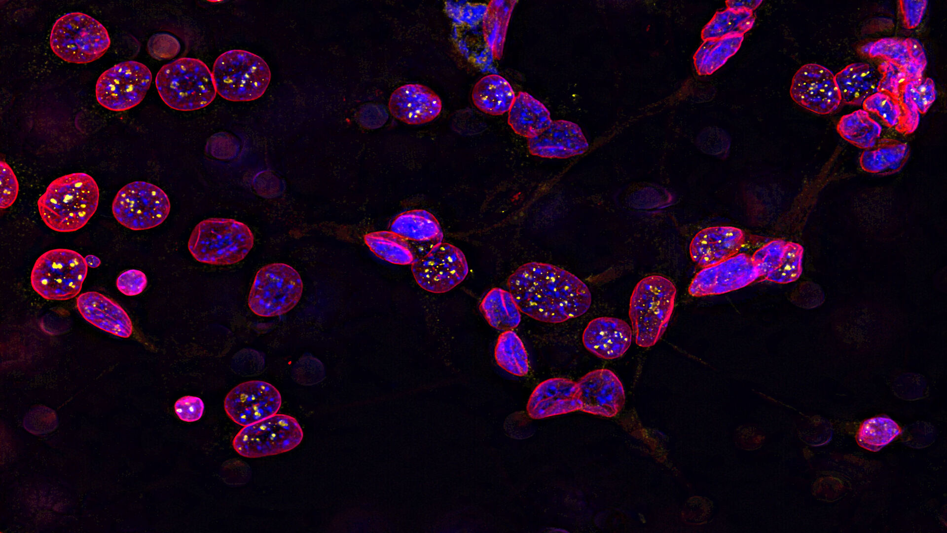
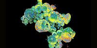
Leica Microsystems unveils next generation STELLARIS with SpectraPlex.

Putting customers first, building partnerships, and adapting to change with a continuous improvement mindset will continue to shape the company's path…

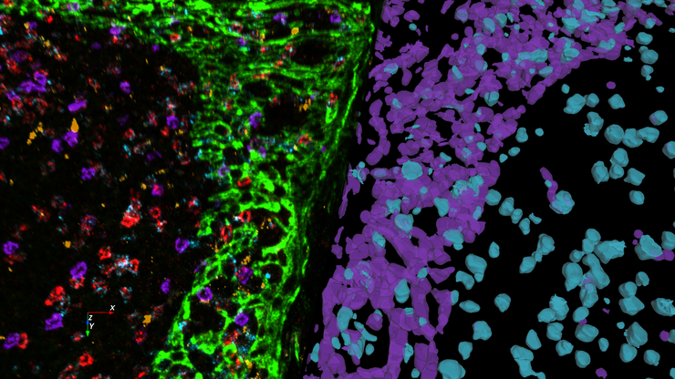
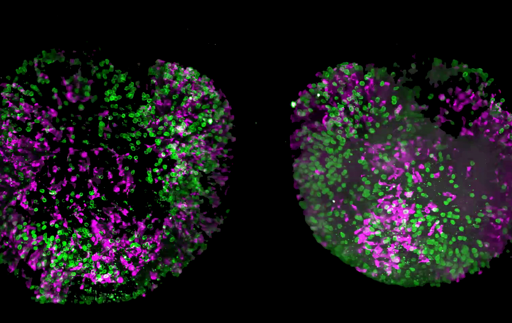
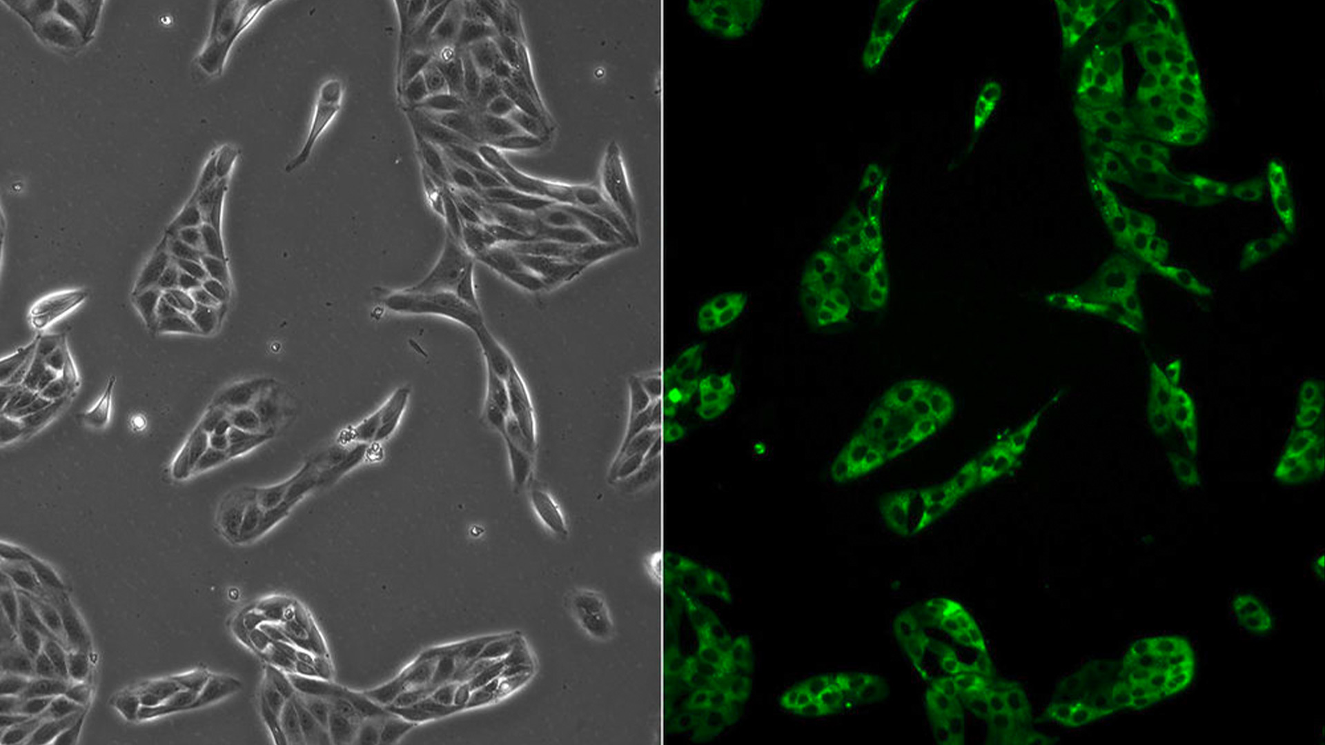
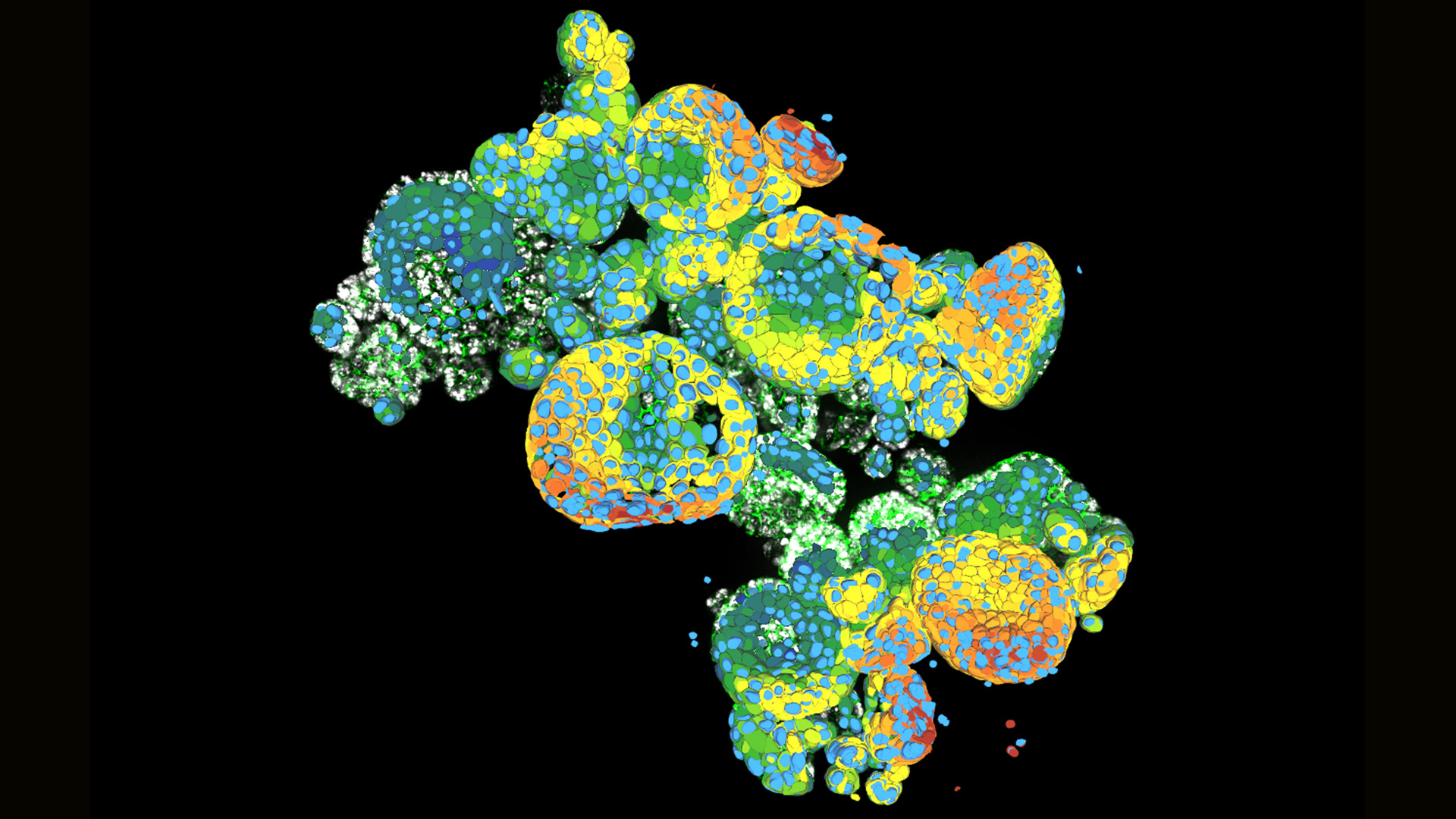
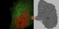
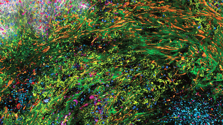
Aivia 14 offers complete multiplexed 3D spatial analysis workflow from cell detection to phenotyping and data exploration

Leica Microsystems announces the official launch of its Partner Excellence Program (PEP) for leading channel partners who outperformed regional growth…

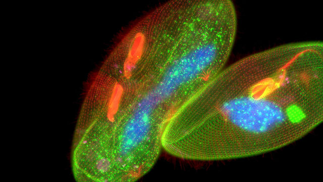
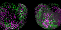
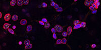
Patented Viventis light sheet solution enables detailed volumetric imaging to explore life in its entirety

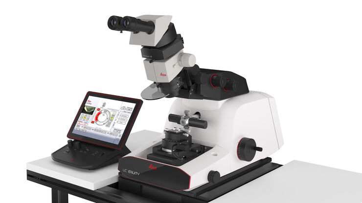
UC Enuity is the next generation ultramicrotome that saves users valuable time through its advanced features and automated-setup functions providing…

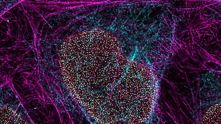
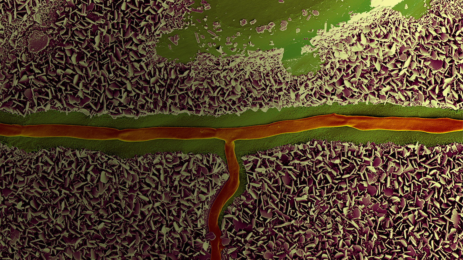
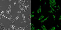
Power to see more with improved resolution and light dose balance

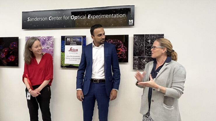
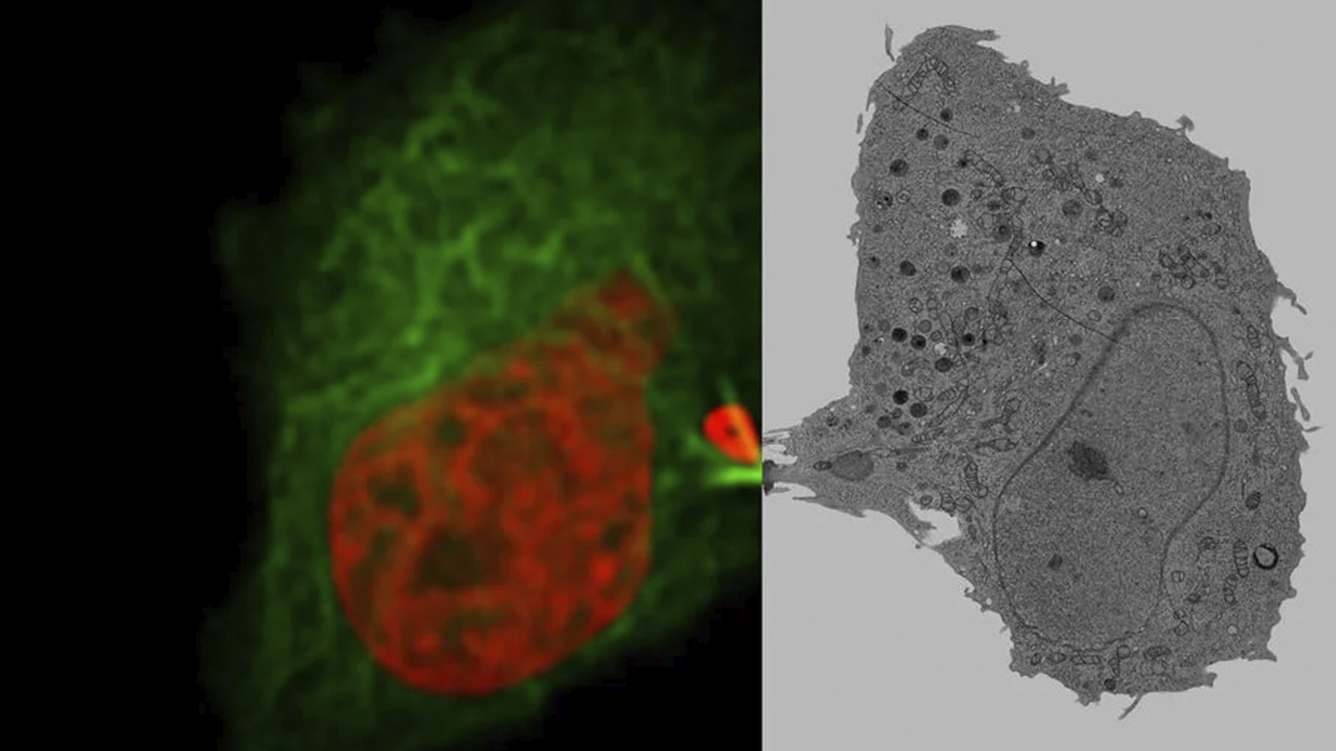
The Sanderson Center for Optical Experimentation (SCOPE) at UMass Chan Medical School and Leica Microsystems, Inc. have collaborated to establish the…

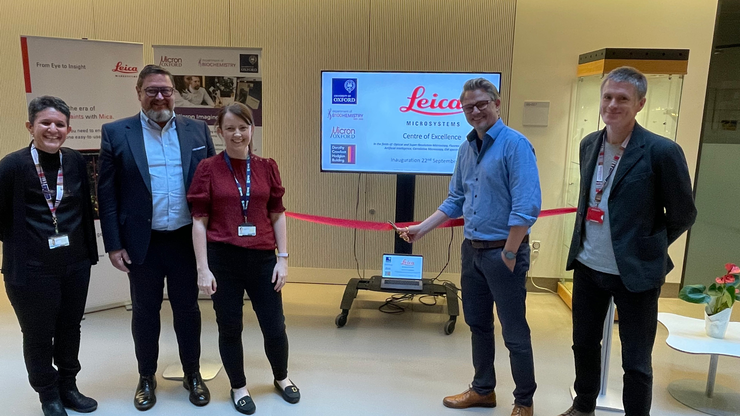
Leica Microsystems has announced an exciting new collaboration with the Department of Biochemistry at The University of Oxford UK, in the fields of…

Scroll to top