6 March 2024, Wetzlar, Germany – Leica Microsystems, a leader in microscopy and scientific instrumentation, has revealed TauSTED Xtend, a new STED microscopy approach that enables extended multicolor live cell imaging at nanoscopic resolution. By combining spatial and lifetime information, TauSTED Xtend resolves details of live or intact specimens at nanoscale. At the same time, the access to that extra level of information allows working at extremely low light dose. This gives scientists more time to perform in-depth studies of biological processes with cutting-edge resolution capabilities.
“TauSTED Xtend ensures that live cell experiments can be carried out over a longer time span thanks to the balance of light exposure and resolution which leads to gentler imaging at stunning nanoscale depths,” said James O’Brien, VP of Life Sciences and Applied Solutions at Leica Microsystems. “Researchers can now reveal the invisible in unexplored areas of research.”
TauSTED Xtend works on-the-fly, allowing researchers to directly monitor fast biological processes at nanoscale resolution. They can therefore make the most of their experimental window by seeing what happens as it happens.
“TauSTED Xtend offers new opportunities for single- and multi-color experiments with green fluorescent proteins and fluorophores that are a workhorse in life science research and currently underused in nanoscopy studies,” said Ulf Schwarz, Application Manager Confocal Microscopy at Leica Microsystems. “Scientists can now effectively scale their experiment down to the nanoscale using familiar protocols and a wide range of fluorescent labels and markers commonly used in biological research.”
TauSTED Xtend is the next step in the evolution of stimulated emission depletion (STED) imaging, offering extended live-cell-imaging times at the cutting edge of nanoscale resolution. Leica developed the proprietary TauSTED approach in 2019, leveraging FLIM and lifetime-based technologies to improve image quality and increase image resolution.
In combination with the Leica STELLARIS 8 FALCON FLIM Microscope, it is now possible to perform multi-fluorophore STED imaging with lifetime-based species separation.
For more information on TauSTED Xtend, read the application note here.
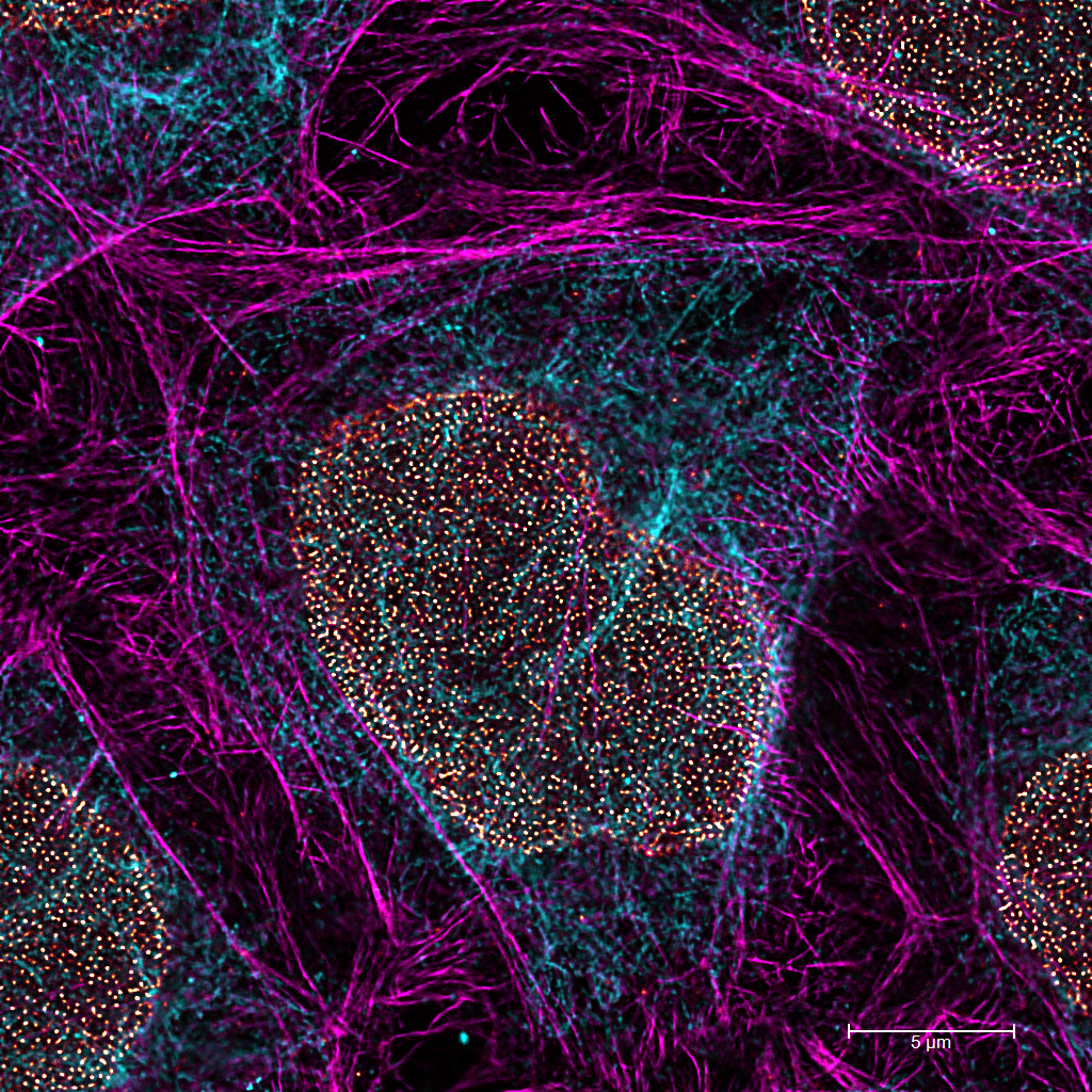
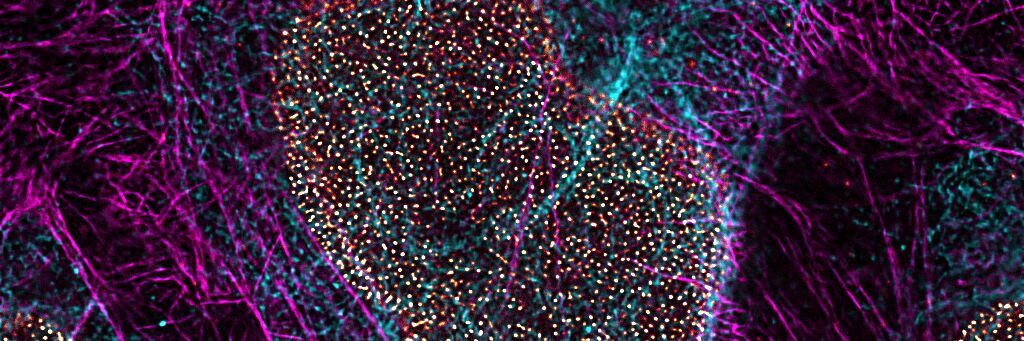
Image caption:
STED for Cell Biology: Multicolor fixed-sample. Image acquired using TauSTED Xtend 775; Vimentin AF 594 (cyan), Phalloidin Multicolor fixed-sample TauSTED Xtend 775; Vimentin AF 594 (cyan), Phalloidin ATTO 647N (magenta) and NUP107 CF680R (glow). Triple color STED imaging with a single depletion line at low STED power. ATTO 647N (magenta) and NUP107 CF680R (glow). Triple color STED imaging with a single depletion line at low STED power. Scale bar: 5 µm.
Sample courtesy of Brigitte Bergner, Mariano Gonzales Pisfil, Steffen Dietzel, Core Facility Bioimaging, Biomedical Center, Ludwig-Maximilians-University, Munich, Germany.
__________________________________________
About Leica Microsystems
Leica Microsystems develops and manufactures microscopes and scientific instruments for the analysis of microstructures and nanostructures. Ever since the company started as a family business in the nineteenth century, its instruments have been widely recognized for their optical precision and innovative technology. It is one of the market leaders in compound and stereo microscopy, digital microscopy, confocal laser scanning microscopy with related imaging systems, electron microscopy sample preparation, and surgical microscopes.
Leica Microsystems has six major plants and product development sites around the world. The company is represented in over 100 countries, has sales and service organizations in 20 countries, and an international network of distribution partners. Its headquarters are located in Wetzlar, Germany.