Meet Mica
The world’s first Microhub
Mica enables microscopy access for all, removes the constraints of traditional four-color fluorescence imaging and radically simplifies workflows.
Live Cell Imaging
Leica Microsystems provides you with intelligent innovations in live-cell imaging. Our solutions help you get the best image quality while protecting your samples.
With the variety of fluorescent proteins and multicolored probes that have been developed, it is now possible to label virtually any molecule. The ability to visualize protein dynamics in vesicles, organelles, cells, and tissues has provided new insights into how cells function in healthy and disease states. These insights include the spatiotemporal dynamics of processes like mitosis, embryonic development, and cytoskeleton changes. When studying live cells, common obstacles include phototoxicity and photodamage. To capture fast biological processes, it is crucial to keep the cells healthy and obtain crisp images for reliable data that are free of artifacts. Live‐cell microscopy often requires a compromise between image quality and cell health. During imaging, certain environmental conditions must be maintained to avoid changes in the cells.
A variety of high-performance Leica live cell imaging solutions can overcome these challenges for live-cell imaging enabling new information for cellular physiology and dynamics to be discovered.
Our experts on solutions for live cell imaging applications are happy to help you with their advice.
Your Live Cell Imaging Needs
To perform successful live-cell imaging experiments, using the right platform is critical. When choosing an optical microscope for live‐cell imaging, the following 3 variables should be considered: detector sensitivity (signal‐to‐noise ratio), specimen viability, and image-acquisition speed.
Methods suitable for live-cell applications enable visualization of the dynamics without causing cell damage, as it can affect the results.
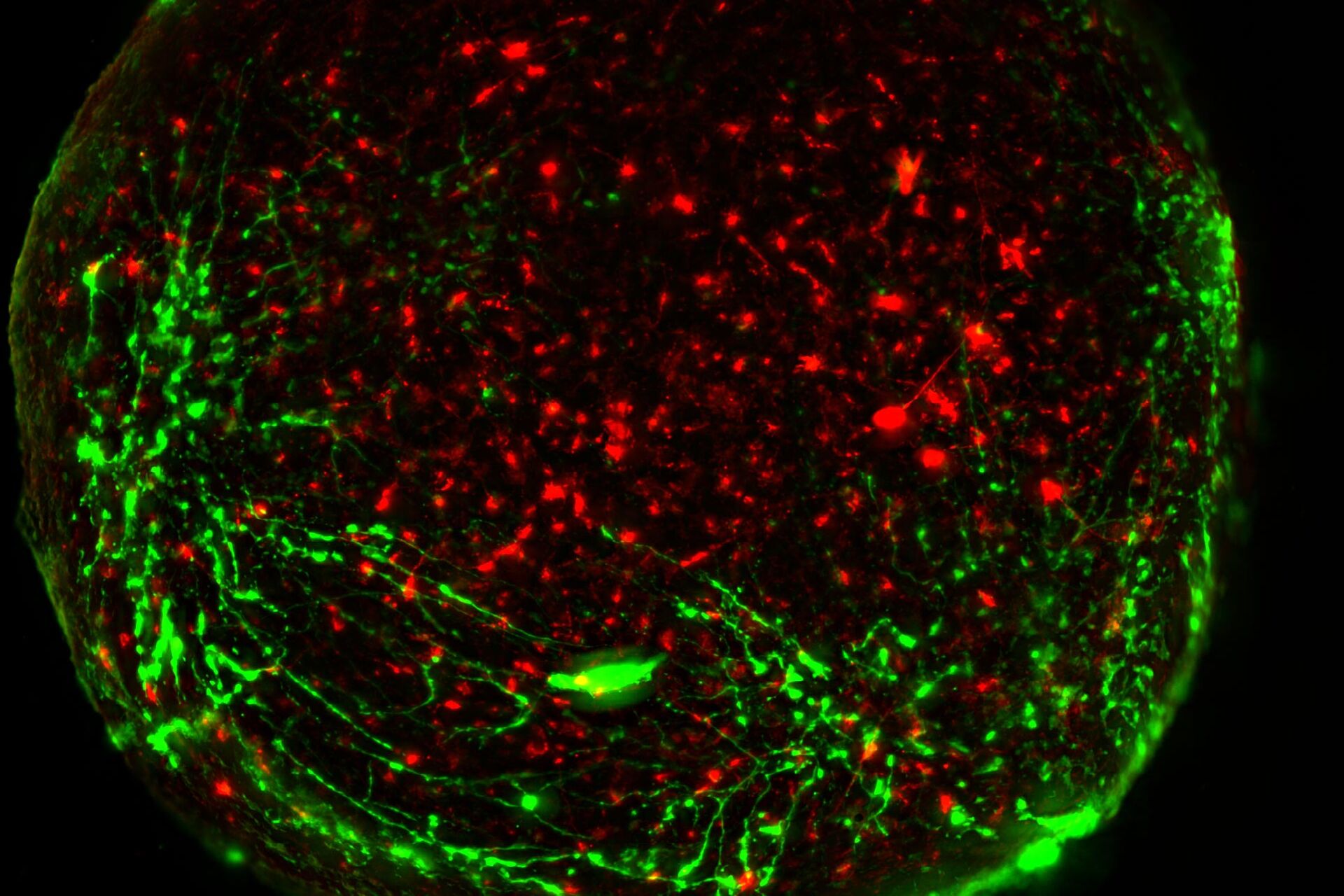
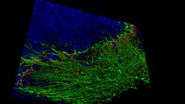
Visualization of brain organoids with a THUNDER Imager Live Cell – Nucleus (DAPI), p-vimentin (AF488), DCX (AF568), PAX6 (AF647). Uncleared human brain organoid (4 colors); Image courtesy by Atria Kavyanifar, M.Sc. (supervised by Prof. Dr. Lie, Prof. Dr. Winner) University Clinic Erlangen, Germany).
Leica Microsystems offers the latest innovations in widefield and confocal imaging technologies for fast 3D live cell imaging with the THUNDER Imagers, Mica – the world’s first Microhub, STELLARIS confocal platform, and FLIM.
Live‐cell imaging is mainly performed with fluorescence microscopy. Widefield microscopy, providing flexible excitation and fast acquisition, is typically used to visualize cell dynamics and development over long times. Confocal microscopy is typically used to study subcellular dynamic events. Multiphoton microscopy allows excitation with longer wavelength light reducing photobleaching and extending cell viability. Finally, fluorescence lifetime imaging (FLIM) can be applied to study fast dynamic signaling events in cells.
Cell Viability & Dynamics During Live Cell Imaging
Leica Microsystems provides you with intelligent innovations in live-cell imaging. Our solutions help you get the best image quality while protecting your samples. Most cellular processes occur in 3 dimensions over time. Therefore, cells need to be imaged in four dimensions (XYZ and time) to obtain a complete picture. Time‐lapse imaging is used to capture cell events over timescales from seconds to months. Repeated imaging of cells at particular points in time is also possible. To protect cell viability during this process, live-cell imaging requires the temperature, pH, and humidity to be kept under control. Light exposure should also be at a minimum to avoid phototoxicity.
Leica Microsystems offers imaging solutions that help optimize your study of live cells, even over long periods of time. They provide the necessary image contrast and resolution to facilitate the analysis of dynamic events. Some Leica systems also enable high-speed imaging and absolute correlated labels without spatiotemporal mismatch between labels of the same time point, so no key cellular events are missed.


Live Cell Imaging System DMi8
The modular DMi8 inverted microscope is the heart of the DMi8 S platform solution. For routine to live cell research, the DMi8 S platform is a complete solution. Whether you need to precisely follow the development of a single cell in a dish, screen through multiple assays, obtain single molecule resolution, or tease out behaviors of complex processes, a DMi8 S system will enable you to see more, see faster, and find the hidden.
Live Cell Imaging Products
Filter by Area of Application

Mica
Mica. Everything you need to enable discoveries, unified in one easy-to-use system. Simultaneous 4-color widefield, confocal resolution, AI supported analysis.

K8
Scientific CMOS camera - For Life Science Imaging Applications and Analysis

STELLARIS DLS
DLS Microscope - Digital Light Sheet

STELLARIS 5 & STELLARIS 8
With the STELLARIS confocal platform, we have re-imagined confocal microscopy to get you closer to the truth.

THUNDER Imager Model Organism
The THUNDER Imager Model Organism allows fast and easy 3D exploration of whole organisms for developmental or molecular biology research.

K5
sCMOS Microscope Camera

THUNDER Imager Live Cell & 3D Assay
THUNDER Imagers provide you with a solution for advanced 3D cell culture assays, whether you want to study stem cells, spheroids, or organoids.

THUNDER Imager Tissue
The THUNDER Imager Tissue allows real-time fluorescence imaging of 3D tissue sections typically used in neuroscience and histology research.

PAULA
Smart Cell Imager

STELLARIS 8 CRS
CRS Microscope - Coherent Raman Scattering Microscope

LAS X Life Science
Software platform for life science applications

DMi8 S Platform
For routine to live cell research

STELLARIS 8 FALCON
FLIM Microscope

Infinity TIRF
Multicolor advanced TIRF module

DMi1
Entry level inverted microscope

Leica DM IL LED
Inverted Laboratory Microscope with LED Illumination
Fast 3D in vivo imaging of Nematostella vectensis (Cnidaria) showing endogenous signal for cilia, nematosomes and clusters of freely circulating cnidocytes (green) and dextran red fluorescence (magenta). Endogenous signals and fluorescence separated by TauGating on HyD S, 340 volumes acquired in 12 min 45 s. Scale bar, 50 μm. Sample courtesy of Anniek Stokkerman and Aissam Ikmi, EMBL Heidelberg.
Live cell imaging with STELLARIS confocal platform
The STELLARIS platform provides you with solutions whether you need long acquisition times for high-resolution 3D reconstructions or the highest frame rates to capture rapid dynamic events.
STELLARIS is a completely re-imagined confocal microscope platform. STELLARIS confocal microscopes can be combined with all Leica modalities, including FLIM, STED, DLS, and CRS. With the STELLARIS confocal platform, we have re-imagined confocal microscopy to get you closer to the truth.

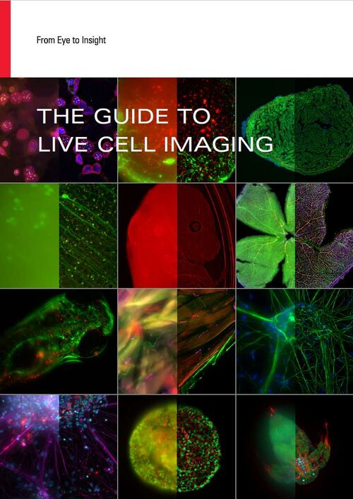
Download The Guide to Live Cell Imaging
In life science research, live cell imaging is an indispensable tool to visualize cells in a state as in vivo as possible.
Live cell imaging related articles

About Live Cell Imaging
Besides the structural organization of cells or organs, dynamic processes are a major contributor to a functioning biological entity. Naturally, these processes can be best observed in living cells with non-invasive techniques like optical methods, collectively called “live-cell imaging” methods. Live-cell imaging covers all techniques where live cells are observed with microscopes – from the observation of embryogenesis with stereo microscopes, via cell growth studies with compound microscopes, until studies of physiological states of cells or cellular transport using fluorescent dyes or proteins. Although being highly demanding for both, experimenter and equipment (e.g. imaging systems, climate control), live-cell imaging techniques deliver results that are indispensable for present-day research.
Interested to know more?
Talk to our experts. We are happy to answer all your questions and concerns.
Contact UsDo you prefer personal consulting?