EM Sample Prep Workflows & Uses
Researchers can consistently achieve high-quality, precise, and reproducible results when imaging samples with electron microscopy by using Leica sample preparation solutions. Our solutions support Scanning Electron Microscopy (SEM), Transmission Electron Microscopy (TEM), and Cryo EM.
Our imaging experts are here to help with advice on solutions for EM Sample Prep Workflows & Uses.

How can researchers prepare samples for SEM?
Sample preparation for SEM (scanning electron microscopy) involves techniques such as coating, drying, and embedding to ensure optimal imaging. Using Leica solutions for SEM samples, researchers improve conductivity, reduce artifacts, and ensure stable, high-quality surfaces for reliable SEM imaging at room temperature.

What methods are best for TEM sample preparation?
TEM (transmission electron microscopy) sample preparation requires ultra-thin, damage-free sections, sputter coating of EM grids, and staining to achieve detailed structure visualization. With the precision and reliability of Leica instruments, researchers can produce high-resolution TEM images.

What is important for cryo-EM sample preparation?
Cryo EM benefits from vitrification by rapid freezing to keep structures in their natural state.
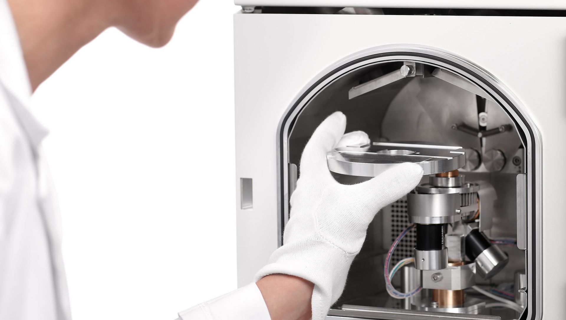
Leica cryo preparation solutions—including advanced vitrification, coating, ultra thin sectioning, cryo planing, and customizable cryo transfer and CLEM workflows—deliver reproducible, contamination free samples. They help maintain native structures, provide precise targeting, and enable high resolution cryo EM imaging.
Why choose Leica instruments and solutions for EM sample preparation?
SEM sample preparation
SEM preparation tools from Leica Microsystems include high vacuum sputter coaters and critical point dryers. These solutions ensure high-quality surface imaging. Plus, Leica instruments provide consistent and reliable results for your SEM studies.
TEM sample preparation
Researchers can rely on TEM preparation solutions from Leica Microsystems for precision and reliability. Essential tools like ultramicrotomes and automated staining systems help produce ultra-thin sections and high-resolution images. Sputter coating enhances sample conductivity and imaging quality.
Comprehensive Cryo-EM preparation
With cryo-preparation instruments from Leica Microsystems, including plunge freezers and high-pressure freezers, researchers ensure rapid freezing and vitrification. Additionally, our sample transfer system for cryo workflows provides transfer options for all kinds of samples. This option protects them from contamination, ensuring accurate and detailed Cryo-EM analysis.
Frequently asked questions about EM Sample Prep Workflows & Uses
Sample preparation for EM involves the following key steps to ensure that samples are stable, appropriately thinned and have proper contrast for high-resolution imaging:
- Fixation to stabilize biological structures using chemical or cryogenic methods
- Removing water or rapidly vitrifying the sample in cryo workflows
- Embedding for ultrathin sectioning or mounting samples on supports or grids
- Producing electron transparent ultrathin sections
- Coating or Staining
- Moving samples into the EM under clean, controlled conditions to avoid contamination or warming.
Leica Microsystems provides solutions for all these steps to deliver reproducible, high quality results from SEM, TEM, and Cryo EM imaging.
Leica solutions support Scanning Electron Microscopy (SEM), Transmission Electron Microscopy (TEM), and Cryo EM, covering both surface and internal structure imaging workflows.
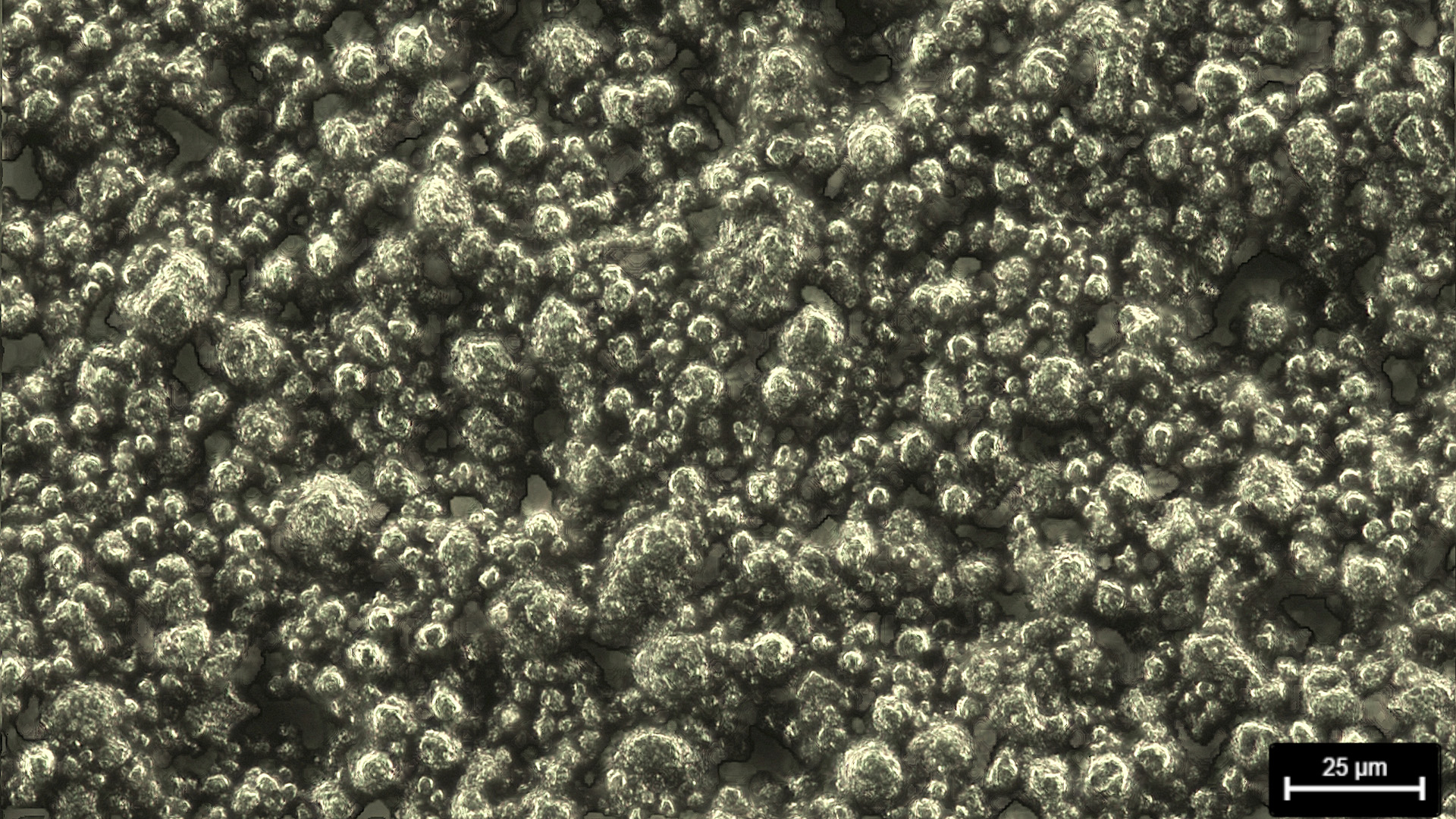
SEM preparation typically involves fixation, dehydration followed by critical point drying to avoid collapse of delicate structures, coating with thin conductive layers, e.g. gold, platinum or carbon to optimize surface imaging. Leica Microsystems provides sputter and carbon coaters, as well as critical point dryers, that help to achieve clean, stable, and conductive surfaces for consistent data quality.
Coating improves conductivity and reduces charging on non conductive samples, which enhances signal to noise and image stability. Sputter coating or carbon coating is selected based on sample type and imaging goals.
Critical point drying removes liquid without surface tension effects, preserving delicate microstructures that might distort or collapse during conventional air drying. It is commonly applied to biological and porous materials intended for SEM.
Transmission Electron Microscopy (TEM) requires electron-transparent samples. Small particles or thin films of <100 nm are sufficiently thin for TEM, but thicker samples are often cut into ultra thin sections by ultramicrotomy. In addition, appropriate staining, and stable supports to visualize internal structures at high resolution. Ultramicrotomes and automated staining systems enable consistent, nanometer scale sections and reliable contrast.
Ultra thin sectioning produces nanometer scale slices of a sample so electrons can transmit with minimal scattering, revealing internal ultrastructure. Precision ultramicrotomy is essential to achieve uniform thickness and reproducible results.
Staining enhances contrast of specific structures by differentially interacting with components of the sample. Automated systems help standardize staining protocols for reliable and comparable TEM datasets.
Cryo EM preserves native structures by rapid freezing and vitrification instead of chemical fixation and dehydration. The workflow maintains cryogenic conditions during transfer and imaging to minimize contamination by ice crystals and structural artifacts.
Plunge freezing vitrifies small or thin samples extremely rapidly, while high pressure freezing is suited for thicker or more complex specimens by suppressing ice crystal formation. Both methods aim to preserve the native state for Cryo EM.
Cryo planing is controlled surface trimming or polishing at cryogenic temperatures to remove contamination, reduce ice artifacts, and expose regions of interest. It helps achieve smoother surfaces for consistent Cryo EM imaging.
Ultra thin cryo sectioning uses cryo ultramicrotomy to produce sections from vitrified samples while maintaining native structures. It enables internal ultrastructure visualization under cryogenic conditions with minimal artifacts.
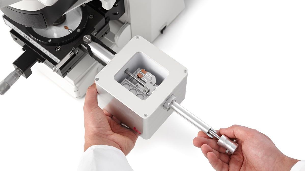
A dedicated cryo transfer system maintains low temperatures, shields samples from environmental exposure, and reduces contamination risk throughout handling and transport, ensuring integrity from vitrification to imaging.
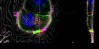
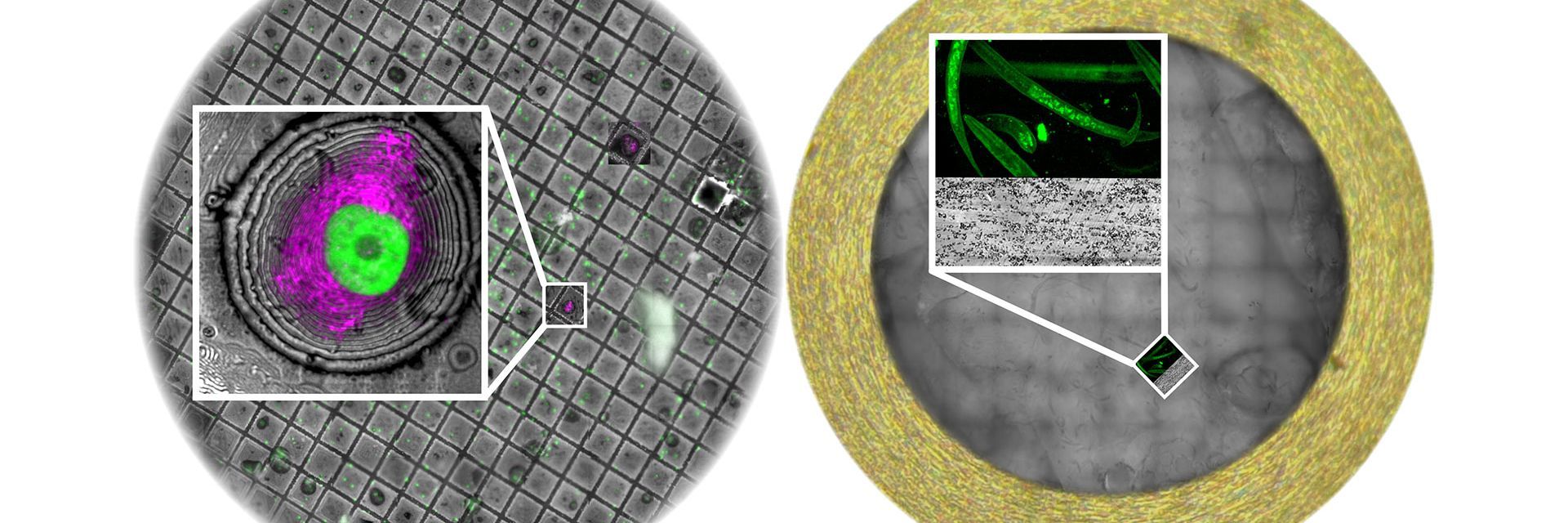
CLEM navigation correlates regions identified in light microscopy (often fluorescence) with the exact coordinates examined in EM. It is valuable when functional or molecular signals from Light Microscopy need to be precisely matched to ultrastructure in EM, including cryogenic workflows.
Choose SEM for detailed surface morphology, TEM for internal ultrastructure at high resolution, and Cryo EM when preserving native state is critical. Your biological question, sample type, and required resolution guide the choice.

 labeled with membrane-permeable calcein, high-pressure frozen in salt water using EM ICE.")
 image of a cross section of C. elegans (roundworm). Courtesy of T. Müller-Reichert, MPI-CBG, Dresden, Germany and K. McDonald, University of California, Berkeley, USA.")
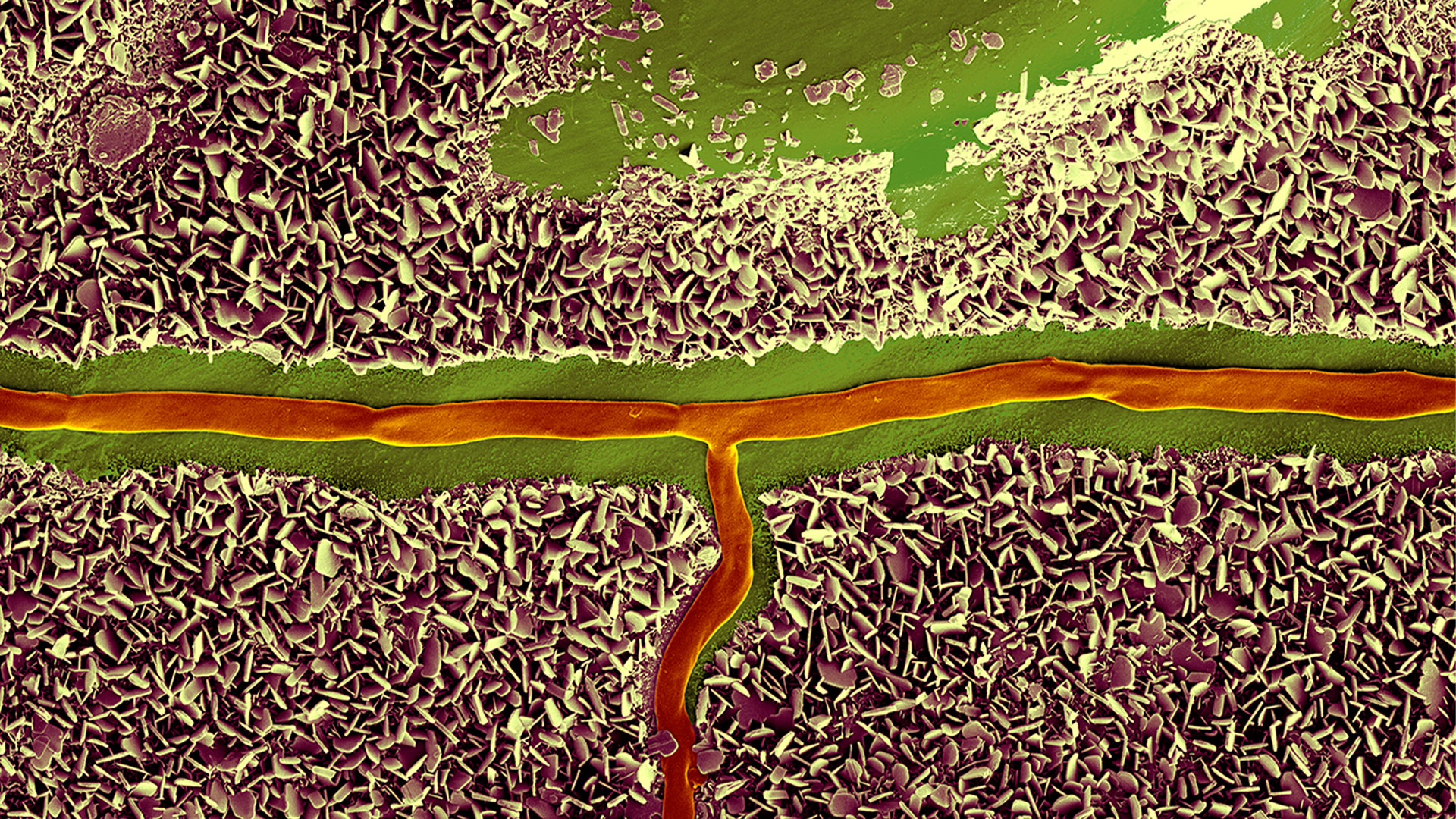
-b-poly(isoprene). Right: Poly(styrene)-b-poly(methyl methacrylate).")


.")