Model organism imaging
Go from overview to fine detail to enhance the understanding of your model organisms with flexible imaging systems from Leica Microsystems.
Leica Microsystems provides innovative microscopy solutions to achieve optimal image quality. These solutions are applicable even with large and complex specimens, such as model organisms, and include platforms for screening and manipulation.
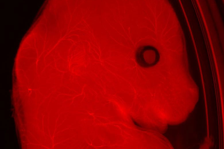

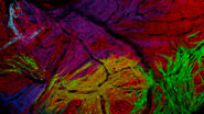
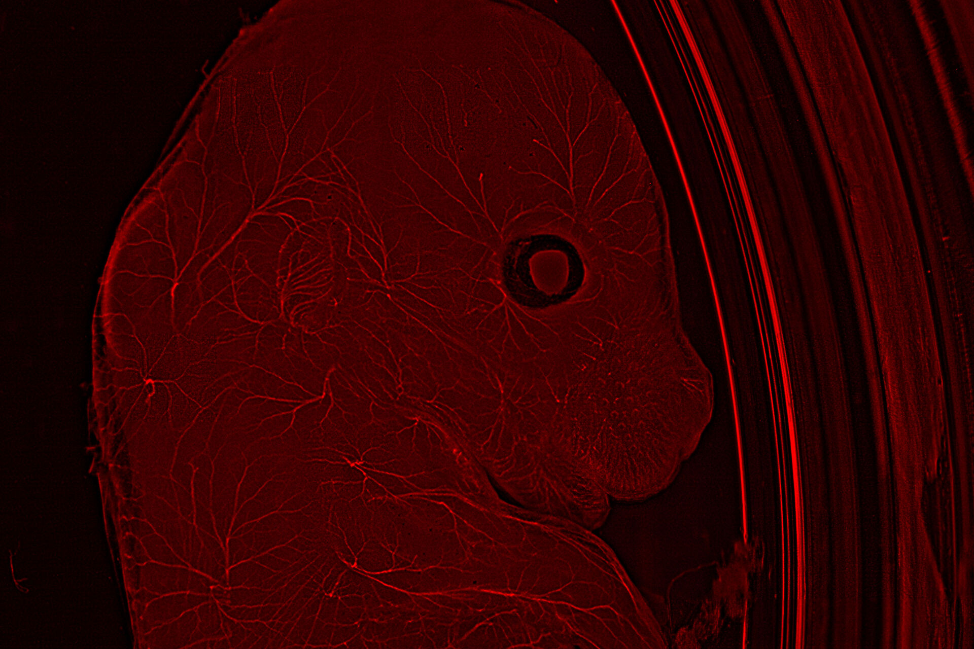
Neurofilaments stained in red to assess neuronal outgrowth in E12-14 mouse. The mouse was uncleared. Courtesy of Yves Lutz, Centre d’imagerie, IGBMC, France.
Microscopy for model organism research
Model organisms are in most cases large, complex, and dense, which can be a challenge. Therefore, there is a need for flexibility from microscope systems to enable the study of these samples. Traditionally, several microscopy techniques are necessary for the study of model organisms, from stereomicroscopes that enable macroscopic analysis and manipulation of specimens such as zebrafish, to multiphoton microscopes that allow deep imaging of large live organisms.
For these thicker specimens, confocal microscopy is the tool of choice to see detail at depth and avoid out-focus-signals. However, it usually poses several challenges, such as slow speeds and photobleaching due to high laser power.
Innovative techniques, such as light sheet microscopy, are becoming more prominent when imaging these light-sensitive samples in 3D thanks to its low phototoxicity.
Related articles
Physiology Image Gallery

How Microscopy Helps the Study of Mechanoceptive and Synaptic Pathways
. Image courtesy of Prof. Hui Guo, School of Life Sciences, Central South University, China")
Imaging Organoid Models to Investigate Brain Health
 acquired using THUNDER Imager Live Cell. Image courtesy of Janina Kaspar and Irene Santisteban, Schäfer Lab, TUM.")

Challenges when imaging model organisms
Work involving model organisms is very heterogeneous and, as such, challenges when using microscopy techniques vary depending on whether they are used for dynamic studies or the study of molecular events, but it can also depend on the complexity of the model organism used, going from the few cells of C. elegans to large complex animals such as the mouse.
A common challenge encountered when imaging model organisms is speed of throughput. Due to the size of the sample, imaging can involve lengthy acquisition times and the analysis of many images to get a complete dataset, which overall reduces experimental efficiency. Speed of acquisition is also an issue when imaging live model organisms, as phototoxicity and photodamage can result in disruption of the sample’s physiology or even cell or sample death. Finally, imaging points within the deep layers of the sample can also produce image artifacts and blurring.