Organoids and 3D Cell Culture
One of the most exciting recent advancements in life science research is the development of 3D cell culture systems, such as organoids, spheroids, or organ-on-a-chip models. A 3D cell culture is an artificial environment in which cells are able to grow and interact with their surroundings in all 3 dimensions. These conditions are similar to how they would be in vivo. Organoids are a type of 3D cell culture containing organ-specific cell types that can exhibit the spatial organization and replicate some functions of the organ.
Organoids recreate a highly physiologically relevant system that allows researchers to investigate complex, multidimensional questions, such as disease onset, tissue regeneration, and interactions between organs. Light microscopy is an essential approach to study the complex interactions and relations with organoids.
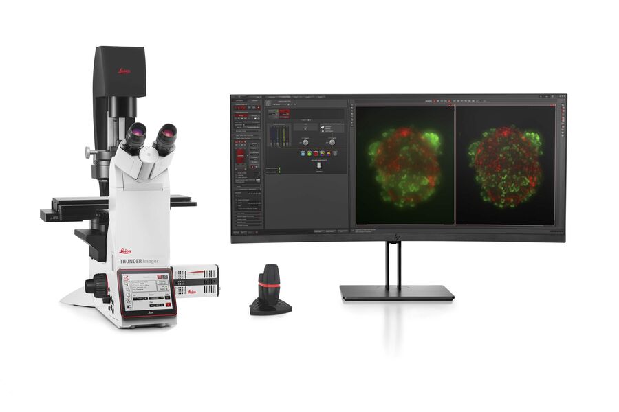
Leica imaging solutions support the study of these multifunctional samples with systems that allow deep and fast imaging for both endpoint measurements or studying the dynamics with live cell imaging.
Our experts on solutions for Organoids and 3D Cell Culture are happy to help you with their advice.
Optimized efficiency for imaging 3D models
Leica Microsystems has developed a number of possibilities that deliver higher performance for the imaging of organoids and other 3D cell culture models.
From traditional methods, such as widefield or confocal microscopy, to more advanced imaging methods, such as multiphoton imaging or LightSheet, Leica Microsystems makes it possible to visualize fine cellular details as well as overall tissue architecture within your 3D cell cultures.
Leica Microsystems offers several solutions for simpler, faster, easier imaging of your 3D cell cultures.
Overcoming challenges in organoid imaging
Imaging is a crucial technique for the study of 3D cell cultures, such as organoids and spheroids.
Effective imaging of organoids poses a new set of challenges as they comprise large volumes. Organoids can be fixed, immunolabeled, and studied using clearing techniques to enable the visualization of their 3D structure. Usually these studies are done using confocal microscopes, as imaging of cultures with more than 2-3 cell layers can be challenging for widefield systems, which have an inherent blur that masks the signal of interest.
Organoids can also be used to study dynamic processes. The study of live organoids faces typical imaging problems, such as phototoxicity and low signal-to-noise ratios, especially when imaging deep in the sample. Recently, fast acquisition microscopy methods, such as FLIM or Lightsheet, have been favored for the study of live organoids as they can be used without altering the physiology of the specimens.
.")


 applied. Image courtesy of Samuel East, Uncommon Bio.")


.")


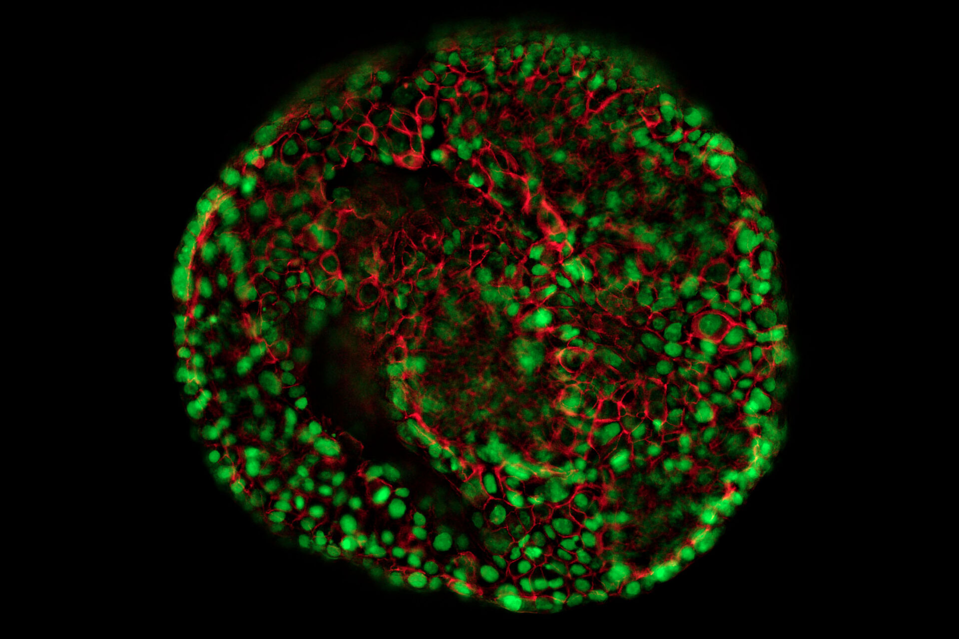
 imaged with the THUNDER Imager 3D Cell Culture. Courtesy of Dr. F.T. Arroso Martins, Tamere University, Finland.")
 acquired using THUNDER Imager Live Cell. Image courtesy of Janina Kaspar and Irene Santisteban, Schäfer Lab, TUM.")
