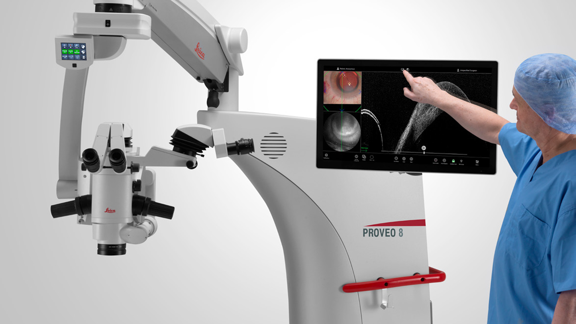


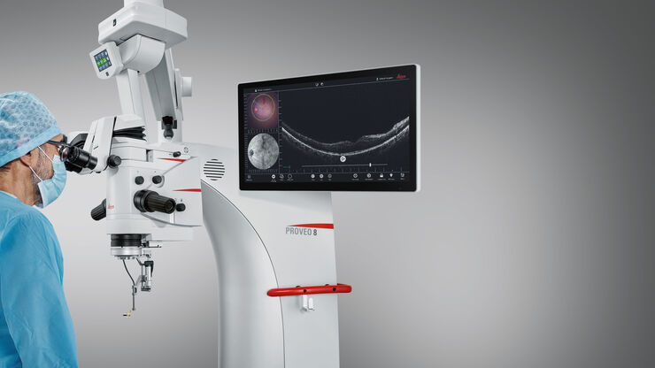
The Proveo 8x 3D digital microscope for ophthalmology debuts at ASCRS 2025 Los Angeles.
Medical Specialties
Medical Specialties
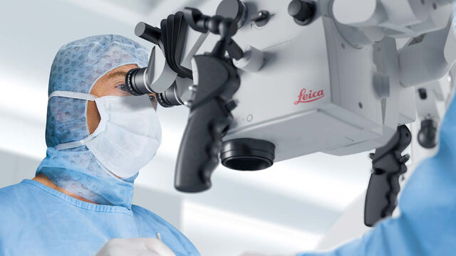
In medical applications, Leica support you with premium optics that deliver crisp, bright, and consistent visualization. To further augment your view, our premium neurosurgery and ophthalmology microscopes are also designed as upgradeable imaging platforms. Easily integrate supplementary digital imaging and recording technologies at any time and ensure you have the visualization you need today, and tomorrow.
In medical applications, Leica support you with premium optics that deliver crisp, bright, and consistent visualization.
To further augment your view, our premium neurosurgery and ophthalmology microscopes are also designed as upgradeable imaging platforms. Easily integrate supplementary digital imaging and recording technologies at any time and ensure you have the visualization you need today, and tomorrow.
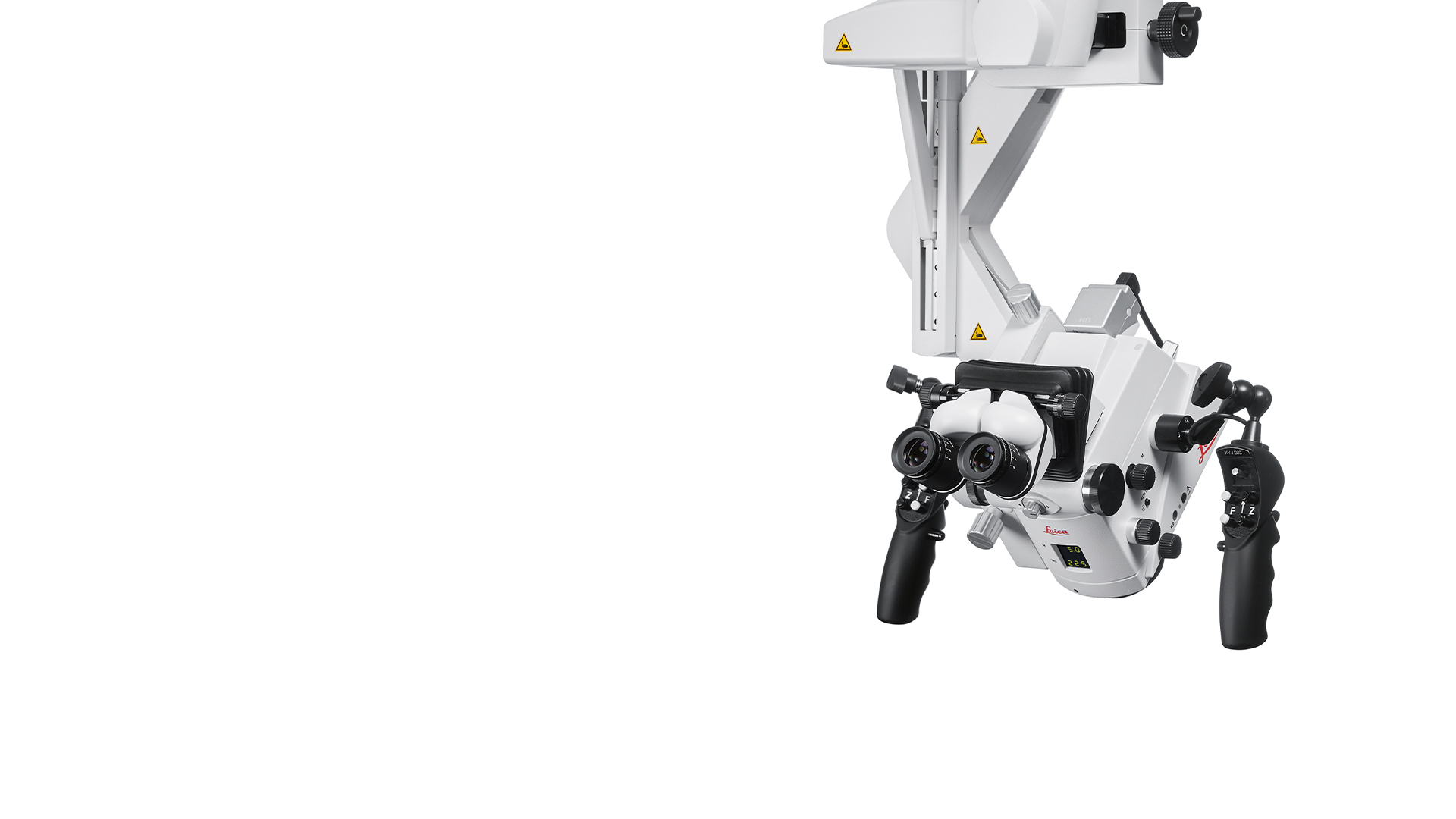
With a portfolio covering microsurgery disciplines including ophthalmology, neurosurgery, otolaryngology (ENT), plastic reconstructive surgery and dentistry, there is a surgical microscope from Leica to meet your needs.
Latest News

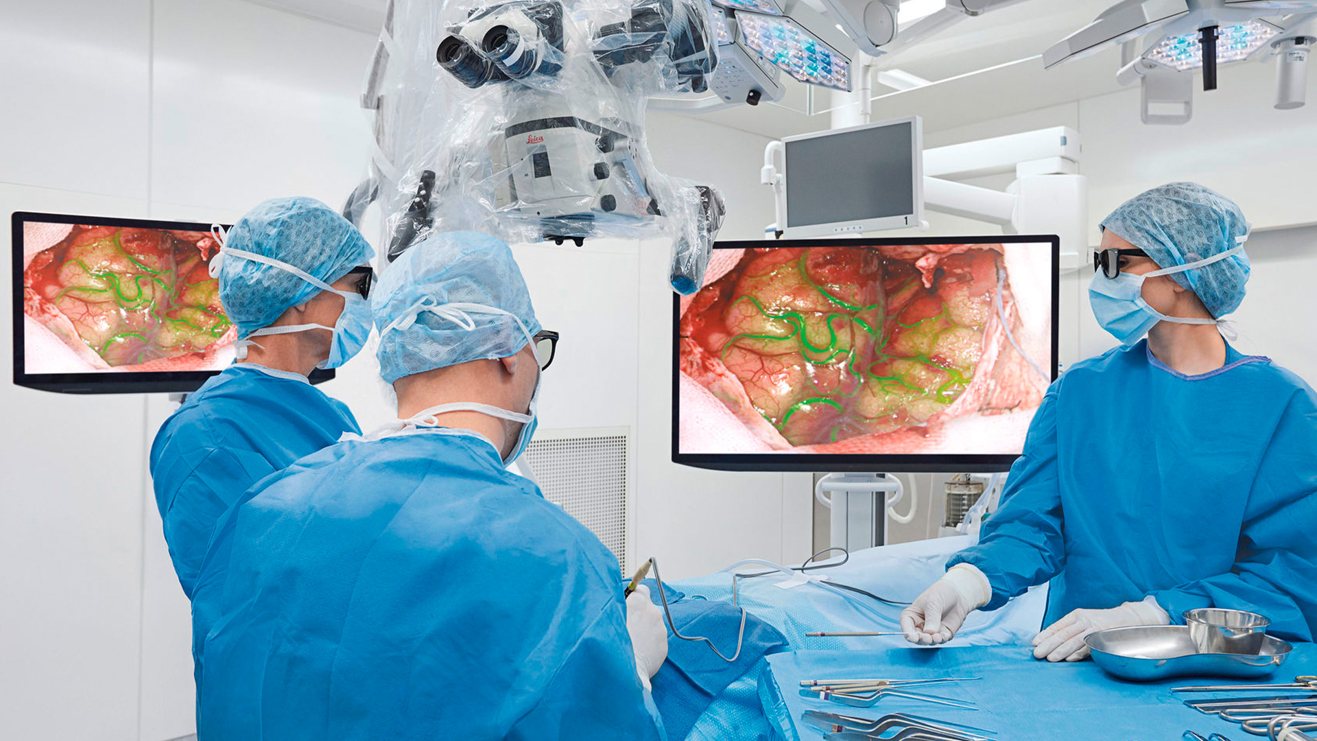
Evolved ARveo 8 surgical microscope leads to clinical value creation for 3D brain-tumor visualization and enables freedom of movement with a surgical…
Next-generation Integrated Intraoperative OCT for Proveo 8 to be Unveiled at the FLORetina ICOOR Meeting 2023
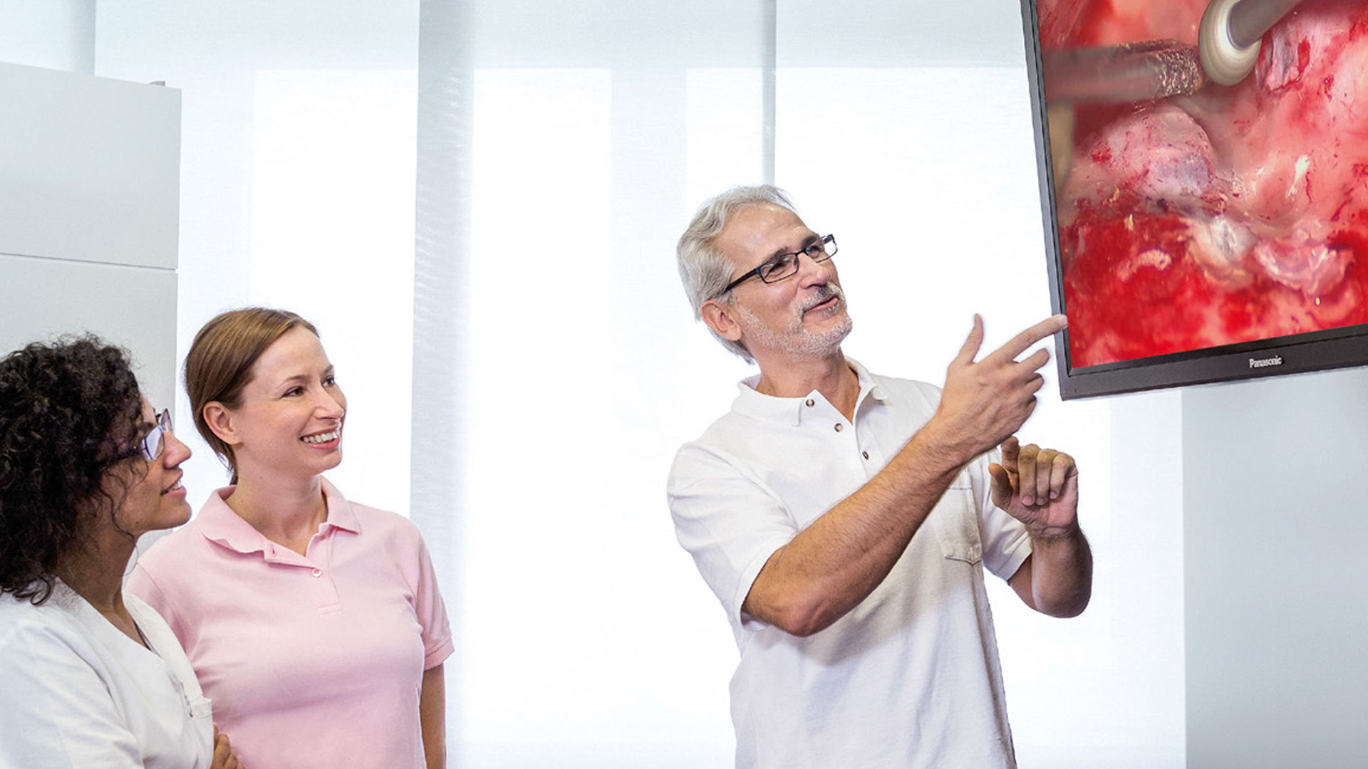
4K camera in ENT microscope supports patient communication
Dental microscope with new integrated 4K camera
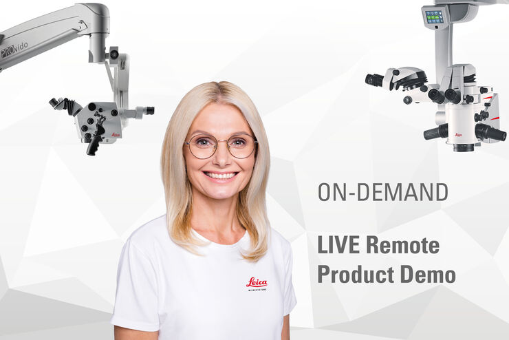
Register now for an easy personal demo of any Leica surgical microscope. Whatever the times, we are there for you, whether on site or online. The…

Interview about the rise of surgical microscopes for use in ENT
Take a look at all our upcoming congresses, exhibitions, webinars, and workshops and join us at one of our next events!
25
–
27
Mar
2026
Congrès SFNC 2026
France
•
24
–
25
Jun
2026
EBME Expo 2026
United Kingdom
•
Congress
