The Orgestra program trains 13 doctoral candidates in developing personalized disease models using stem cell-derived organoids for rare diseases, such as cystic fibrosis and cystinosis. The program is coordinated by Utrecht University and spans 14 partners across seven European countries, offering interdisciplinary and intersectoral training concerning organoid technologies for disease modelling as well as drug discovery and development.
Highlights
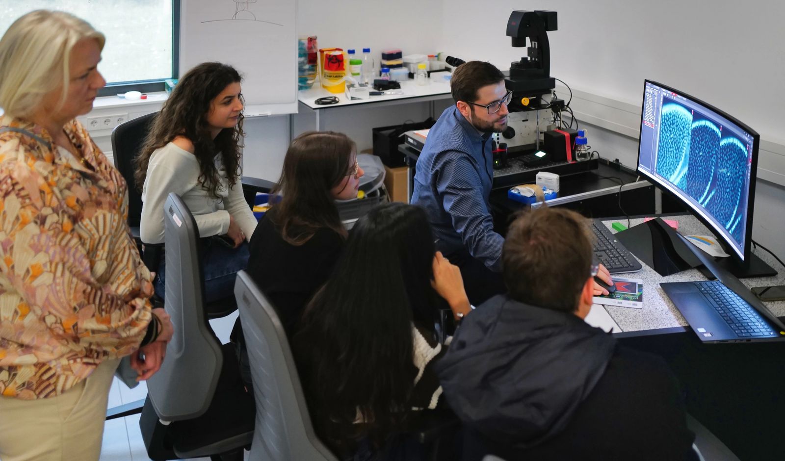
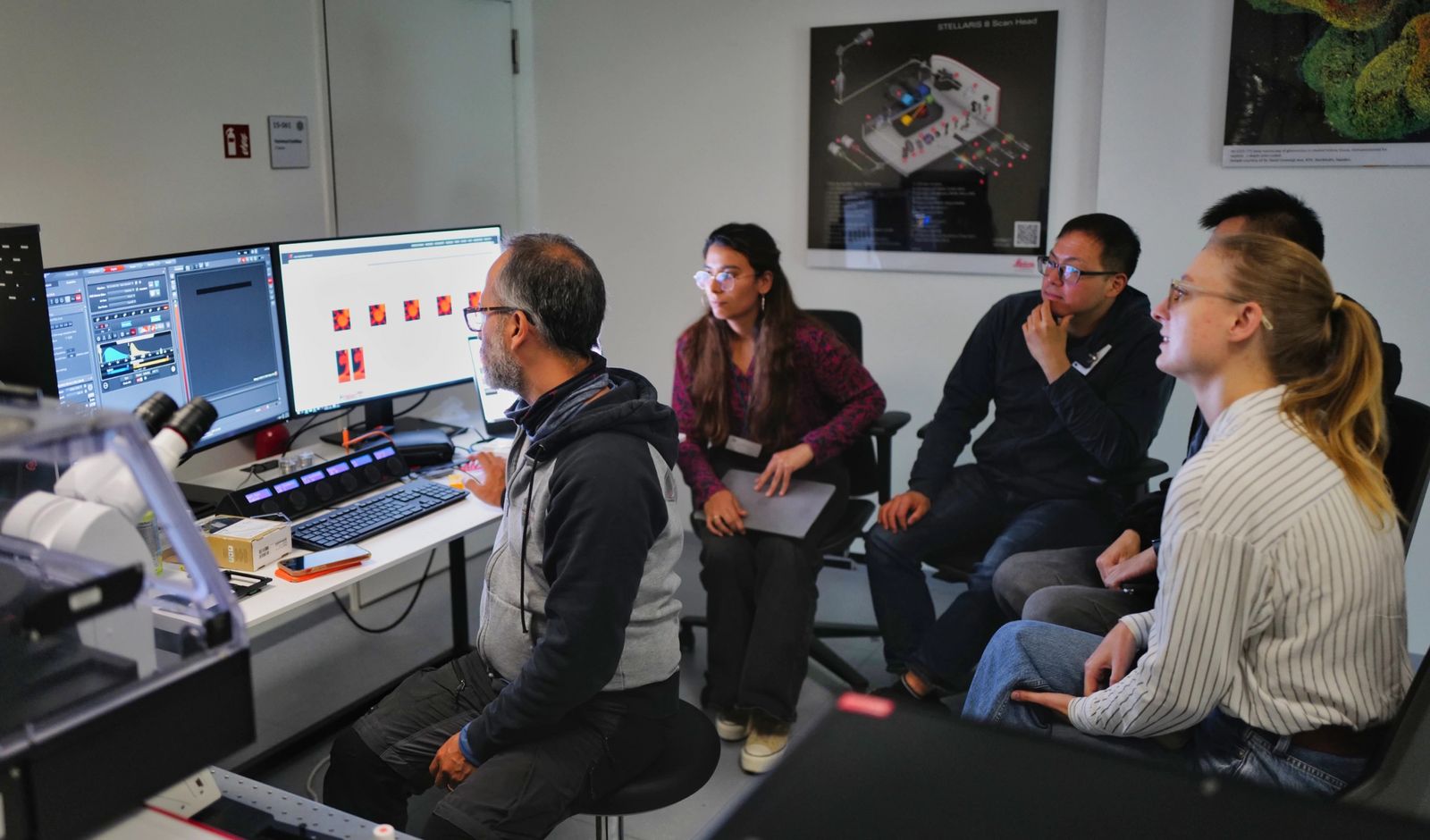
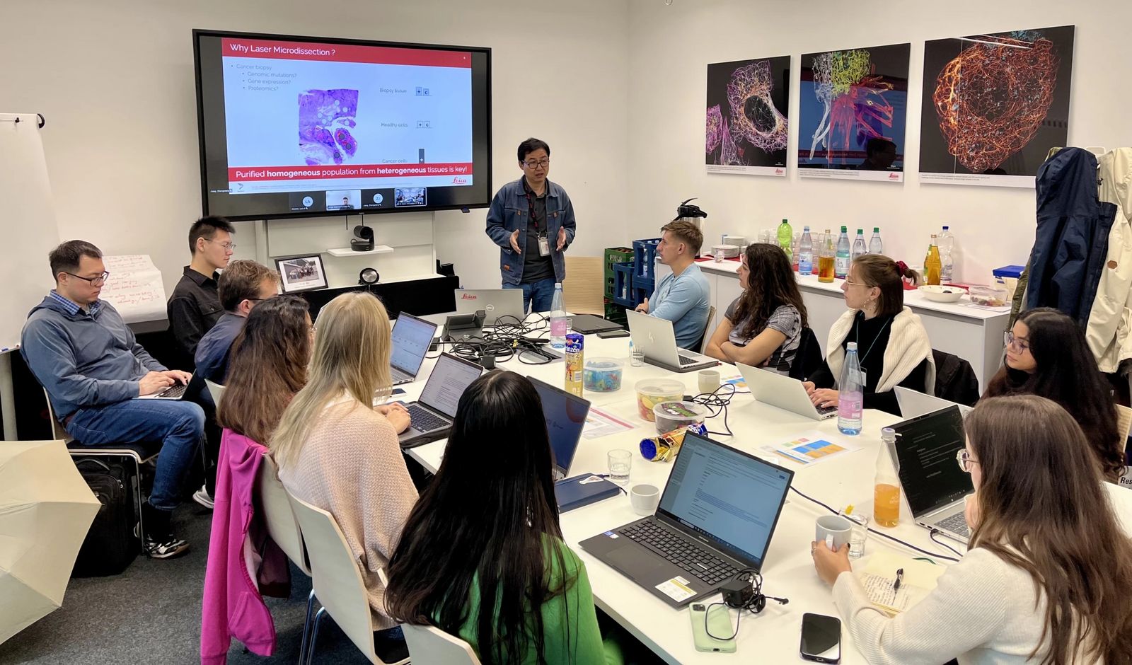
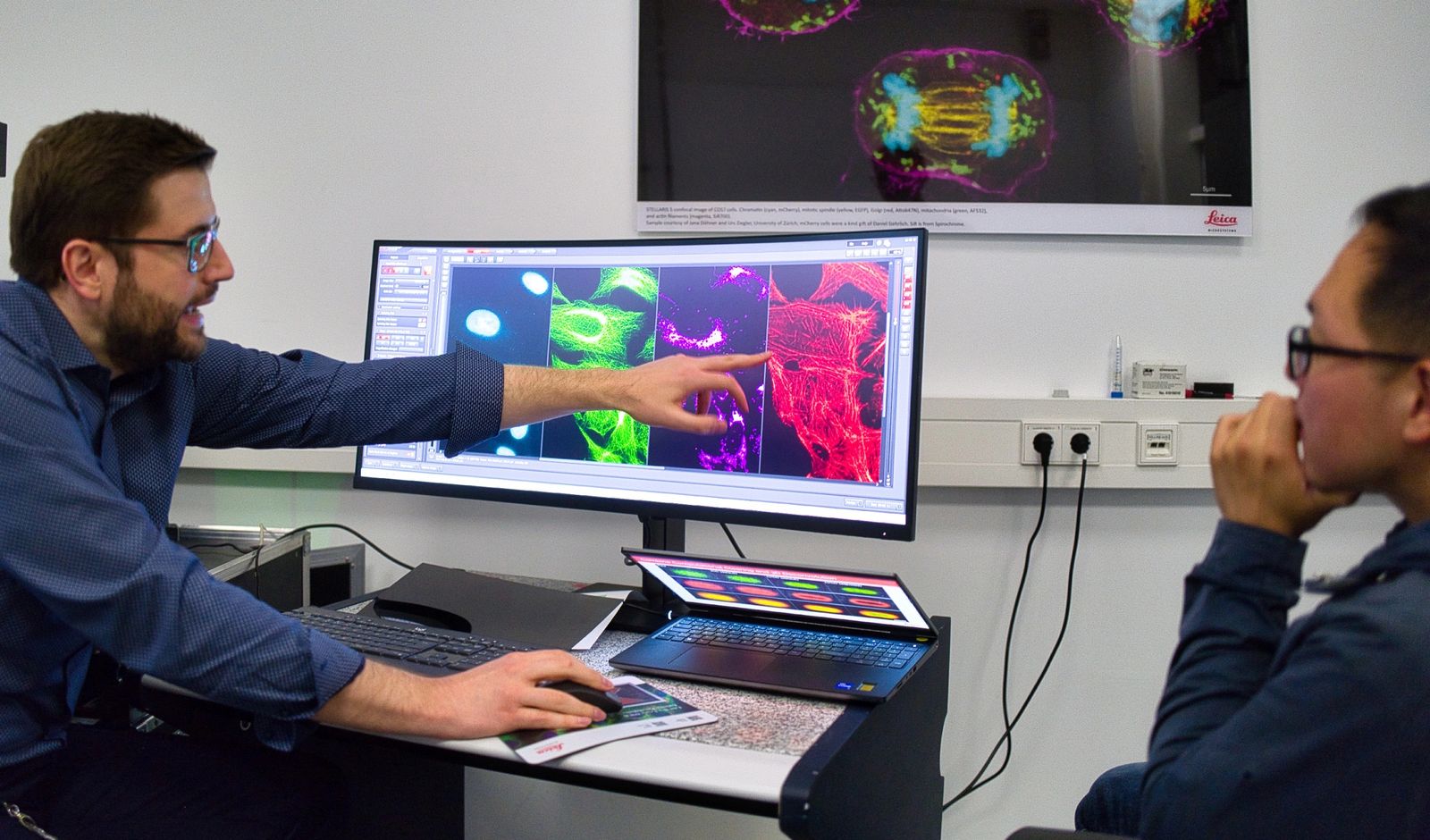
The two-day training focused on microscopy methods and 3D imaging applications, combining lectures with hands-on microscopy sessions. Participants explored a range of advanced imaging techniques including:
- Widefield Microscopy with THUNDER
- Laser Microdissection with LMD7
- Confocal Microscopy with STELLARIS and THUNDER Spinning Disc
- Light Sheet Microscopy with Viventis Deep
- Multi-Photon Microscopy with STELLARIS DIVE
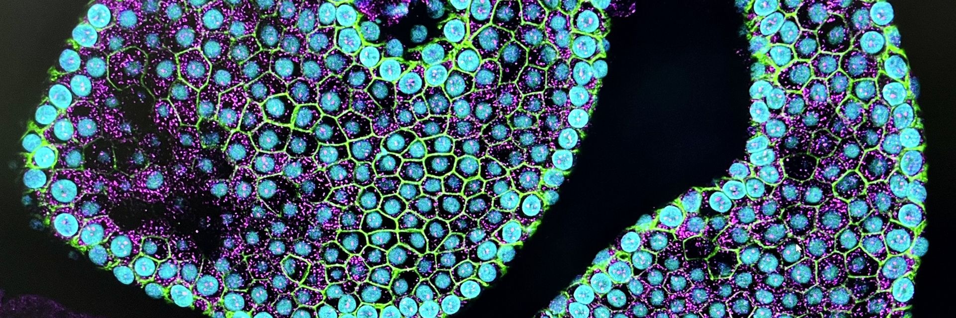
The practical sessions emphasized 3D imaging in complex biological models, such as tubuloids, spheroids, organoids, and 3D cell cultures. Students learned how to optimize imaging parameters and sample preparation strategies tailored to specific scientific questions — a crucial skill for high-resolution visualization and analysis in the field of biomedical research.
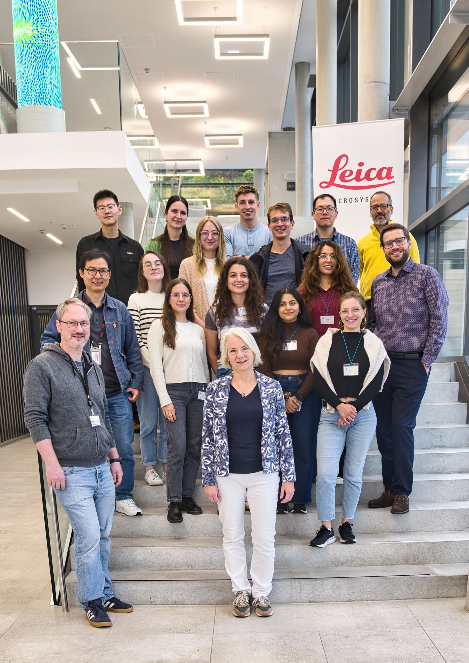
A special thanks goes to our expert trainers, Luis Alvarez, Falco Krüger and Zhongxiang Jiang, whose guidance was instrumental in making the course a success. We also extend our gratitude to the organizers, Wernher Fouquet, Falco Krüger and Roos Masereeuw, for their excellent coordination and support throughout the event.
Looking ahead
We’re excited to welcome two Orgestra PhD students for one-month internships at Leica Microsystems in early 2026. These in-depth placements will allow them to further explore advanced imaging technologies and deepen their expertise in microscopy.
Thank you
We sincerely thank all participants for their enthusiasm, curiosity, and the vibrant atmosphere they brought to the training. It was a pleasure to showcase our advanced imaging portfolio and contribute to the development of the next generation of imaging scientists.