
Industrial
Industrial
Sumérjase en artículos detallados y seminarios web centrados en la inspección eficaz, los flujos de trabajo optimizados y la comodidad ergonómica en contextos industriales y patológicos. Los temas tratados incluyen el control de calidad, el análisis de materiales y la microscopía en patología, entre muchos otros. Este es el lugar donde obtendrá información valiosa sobre el uso de tecnologías de vanguardia para mejorar la precisión y la eficacia de los procesos de fabricación, así como el diagnóstico y la investigación patológicos precisos.

Investigación con Pez Cebra
Para obtener los mejores resultados en clasificación, selección, manipulación y captura y procesamiento de imágenes necesita ver los detalles y estructuras para poder tomar las decisiones adecuadas…

Improving Zebrafish-Embryo Screening with Fast, High-Contrast Imaging
Discover from this article how screening of transgenic zebrafish embryos is boosted with high-speed, high-contrast imaging using the DM6 B microscope, ensuring accurate targeting for developmental…

Overcoming Challenges with Microscopy when Imaging Moving Zebrafish Larvae
Zebrafish is a valuable model organism with many beneficial traits. However, imaging a full organism poses challenges as it is not stationary. Here, this case study shows how zebrafish larvae can be…
How to Radically Simplify Workflows in Your Imaging Facility
VIDEO ON DEMAND - How to radically simplify imaging workflows and generate meaningful results with less time and effort using a highly automated microscope that unites widefield and confocal imaging.

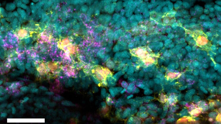
Imaging of Cardiac Tissue Regeneration in Zebrafish
Learn how to image cardiac tissue regeneration in zebrafish focusing on cell proliferation and response during recovery with Laura Peces-Barba Castaño from the Max Planck Institute.

Using U-Shaped Glass Capillaries for Sample Mounting
The DLS microscope system from Leica Microsystems is an innovative concept which integrates the Light Sheet Microscopy technology into the confocal platform. Due to its unique optical architecture,…

Imaging and Analyzing Zebrafish, Medaka, and Xenopus
Discover how to image and analyze zebrafish, medaka, and Xenopus frog model organisms efficiently with a microscope for developmental biology applications from this article.
